
Currículum
Tema 06. Micosis cutáneas
TEMA 06. MICOSIS CUTÁNEAS
0/0
1. INTRODUCCIÓN
0/2

2. DERMATOFITOSIS O TIÑAS
0/8
3. CANDIDIASIS
0/10
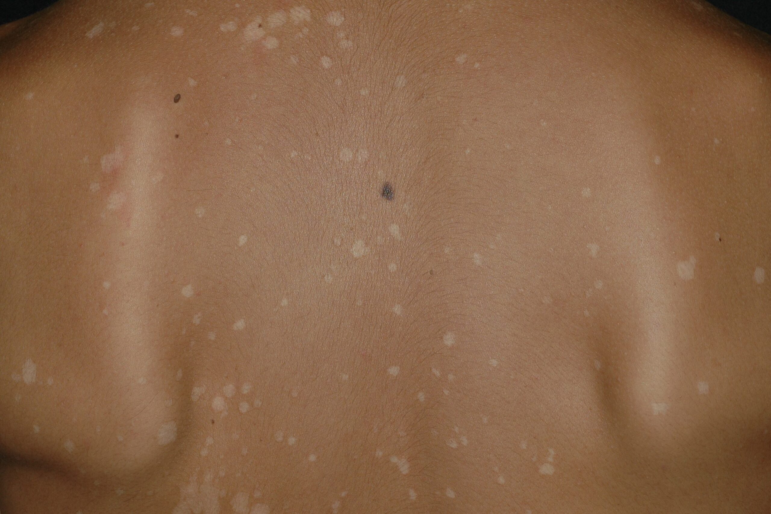
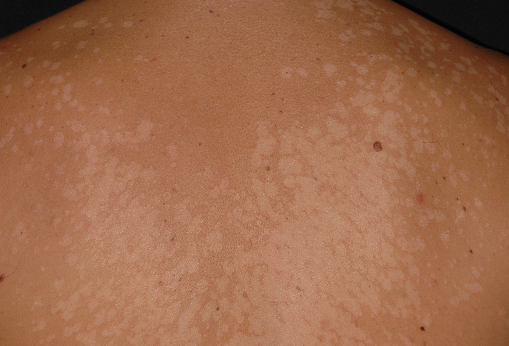
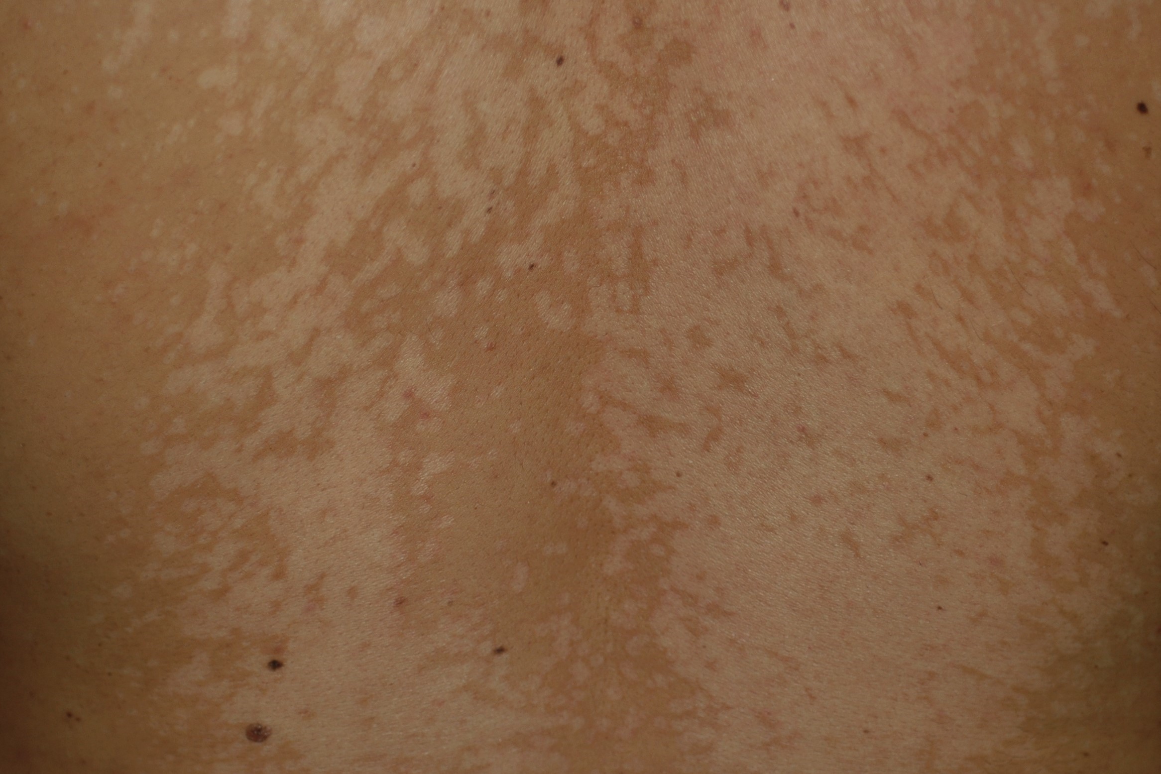
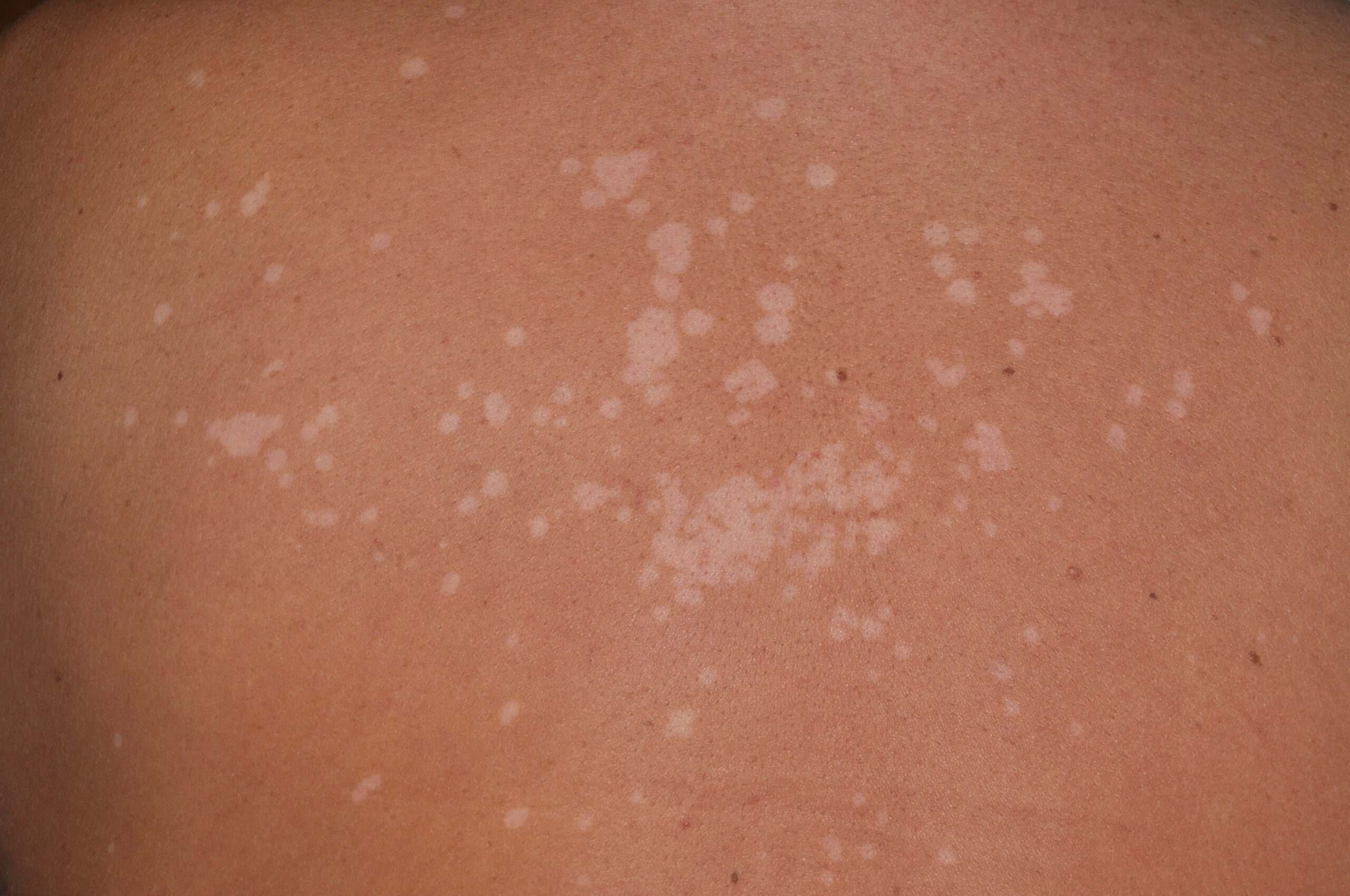
4. PITIRIASIS VERSICOLOR
0/7
5. BIBLIOGRAFIA
0/1
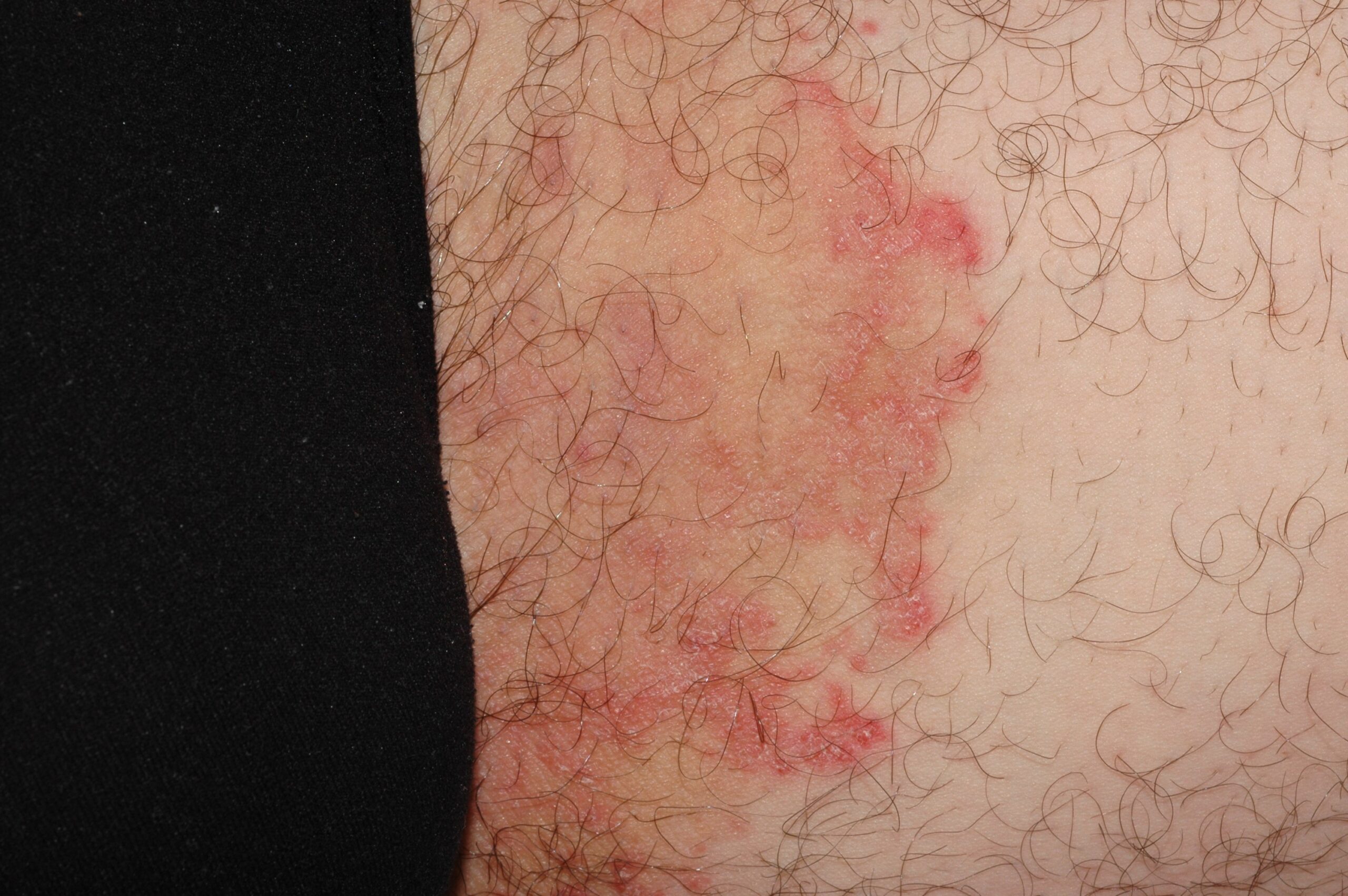
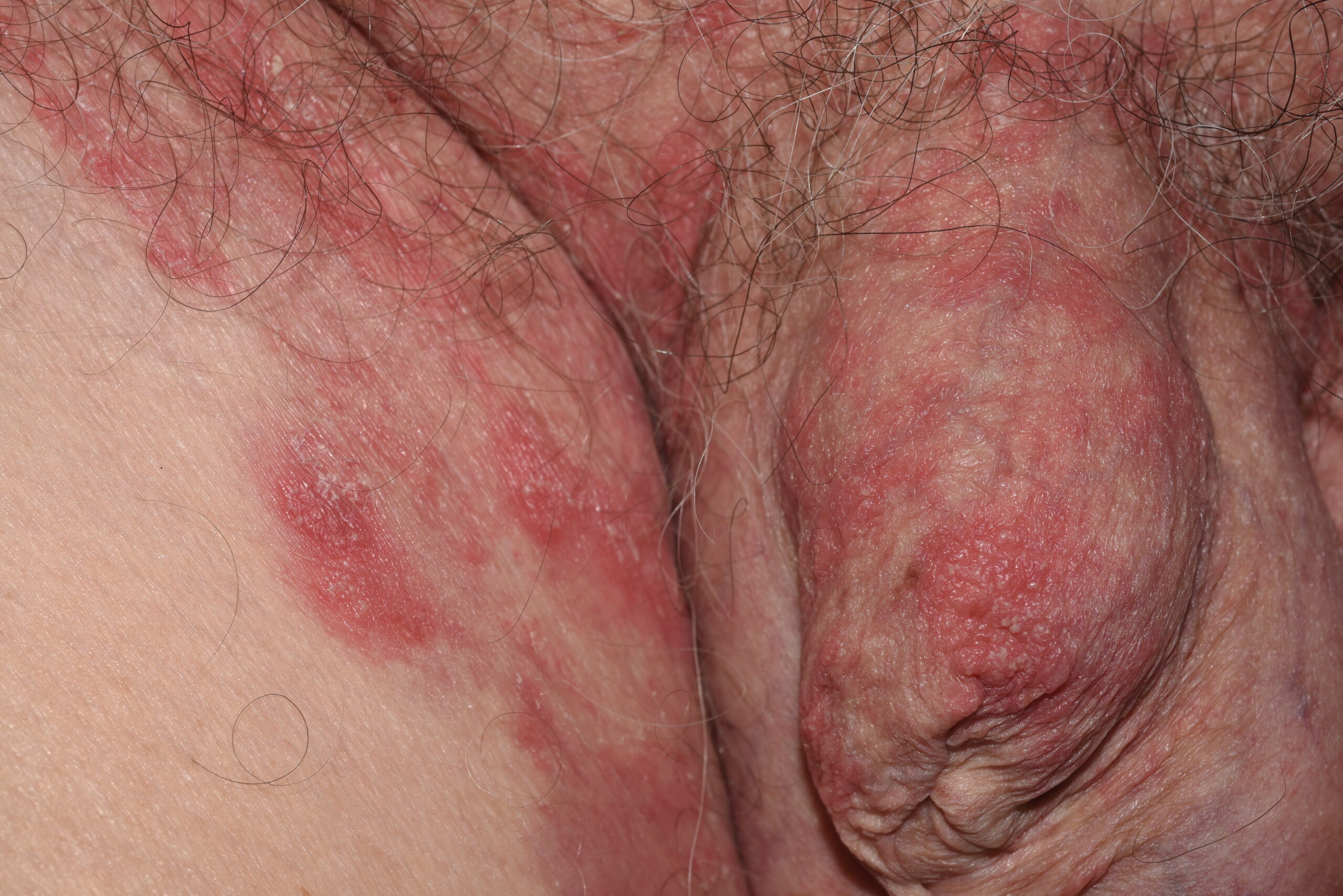
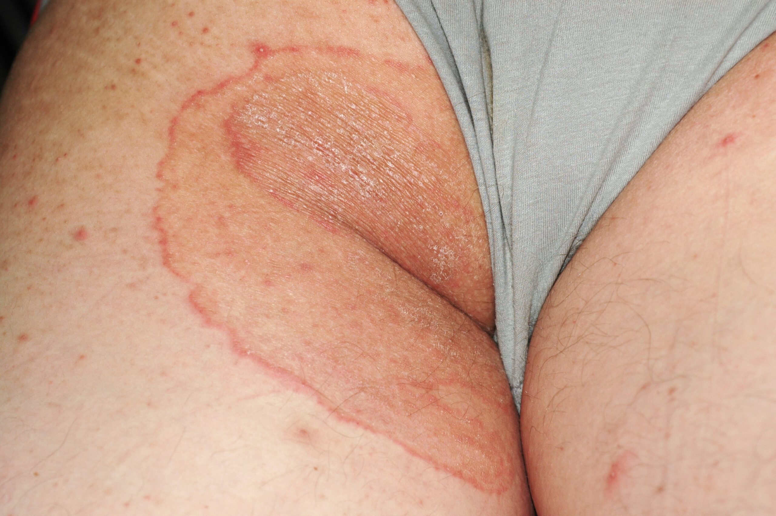
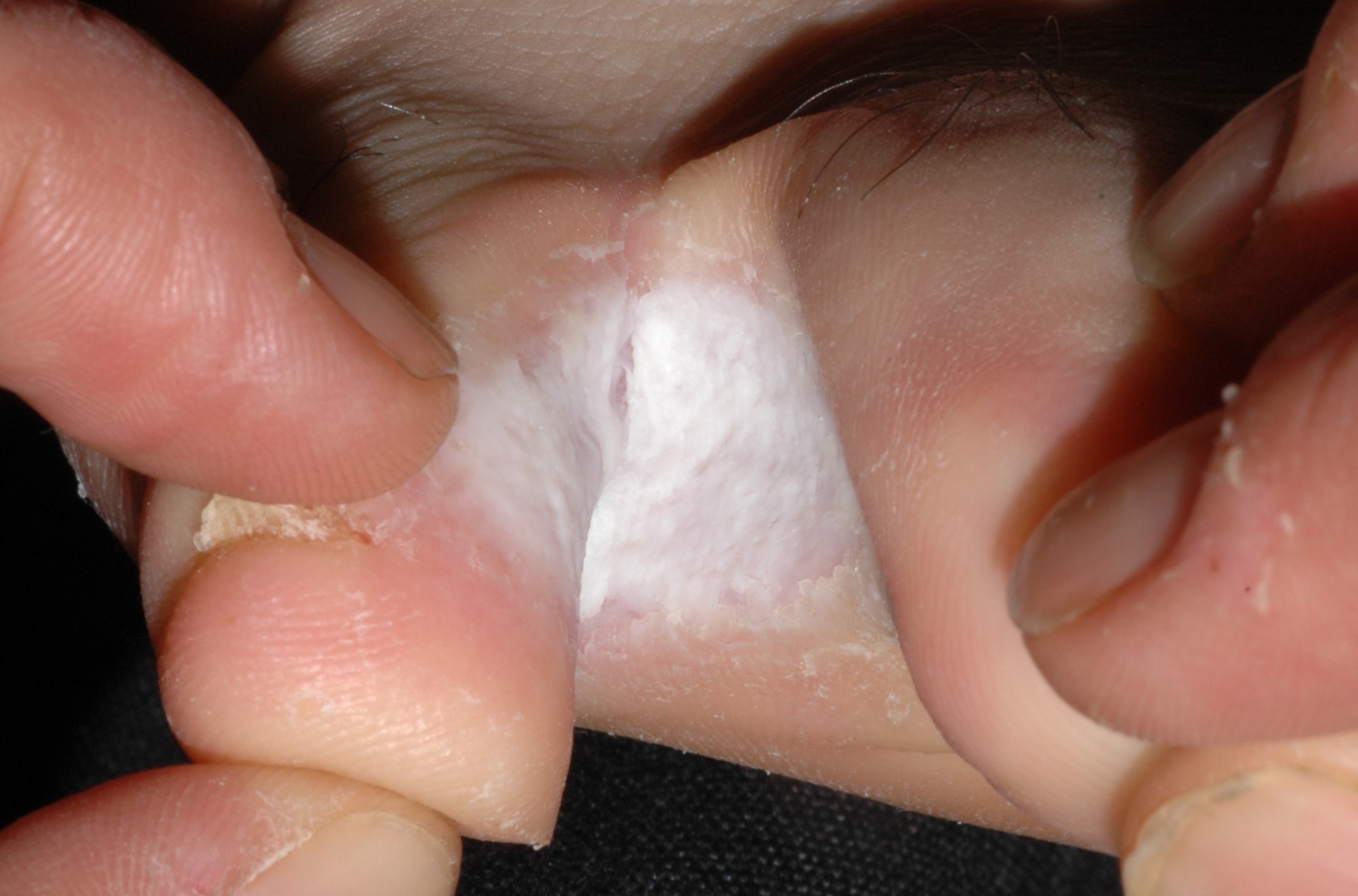
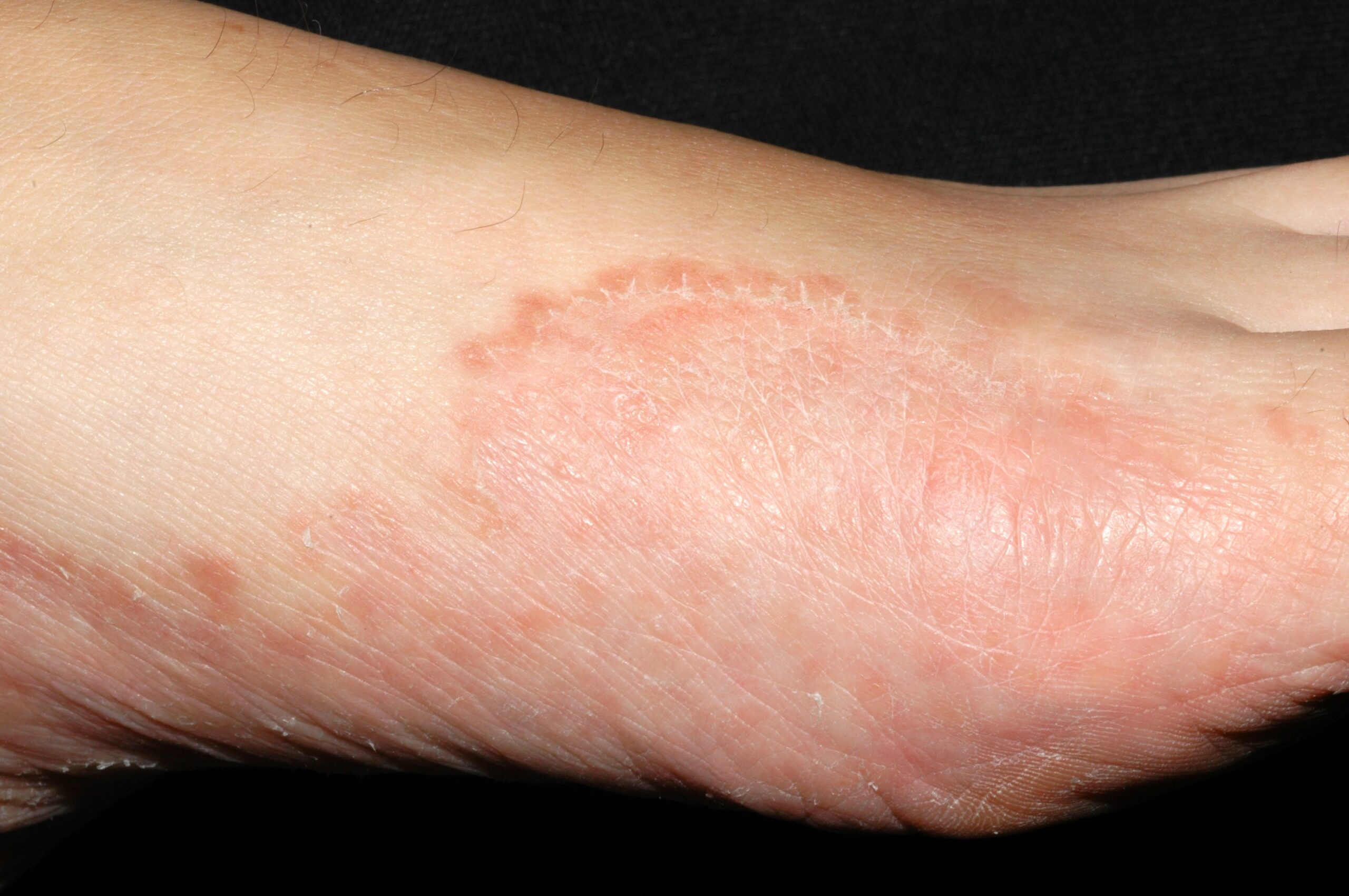
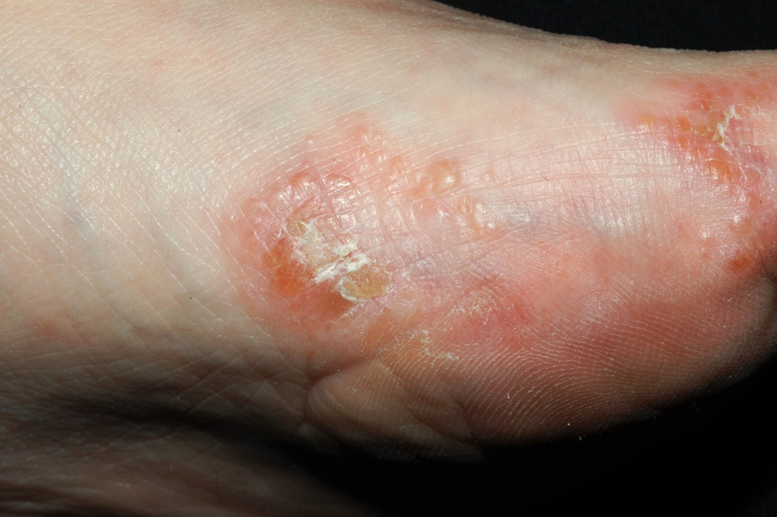
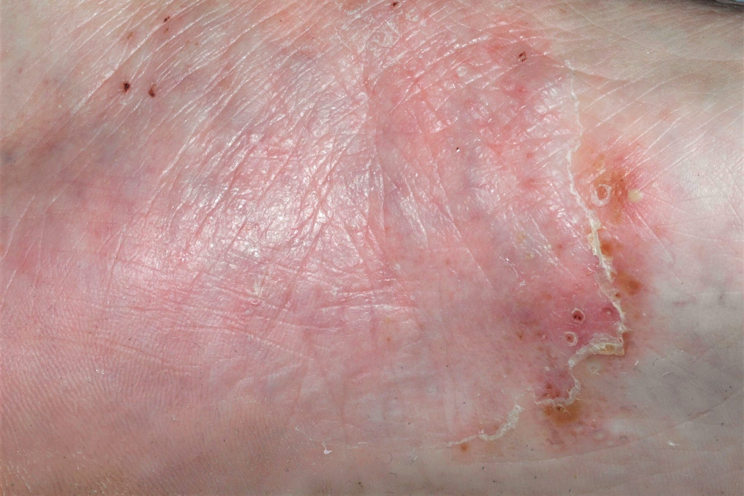
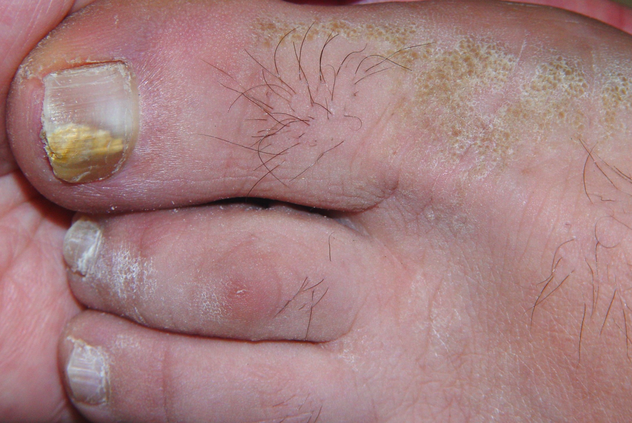
Imágenes tema

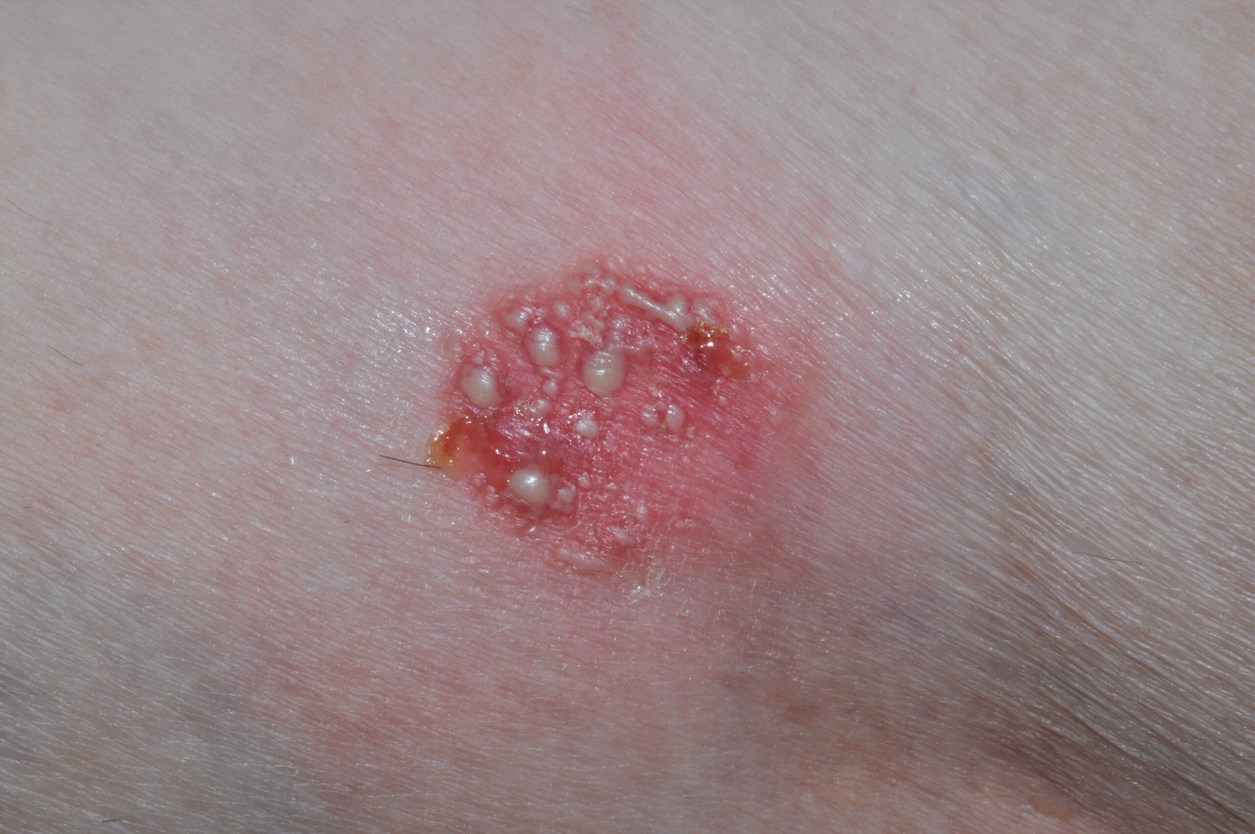
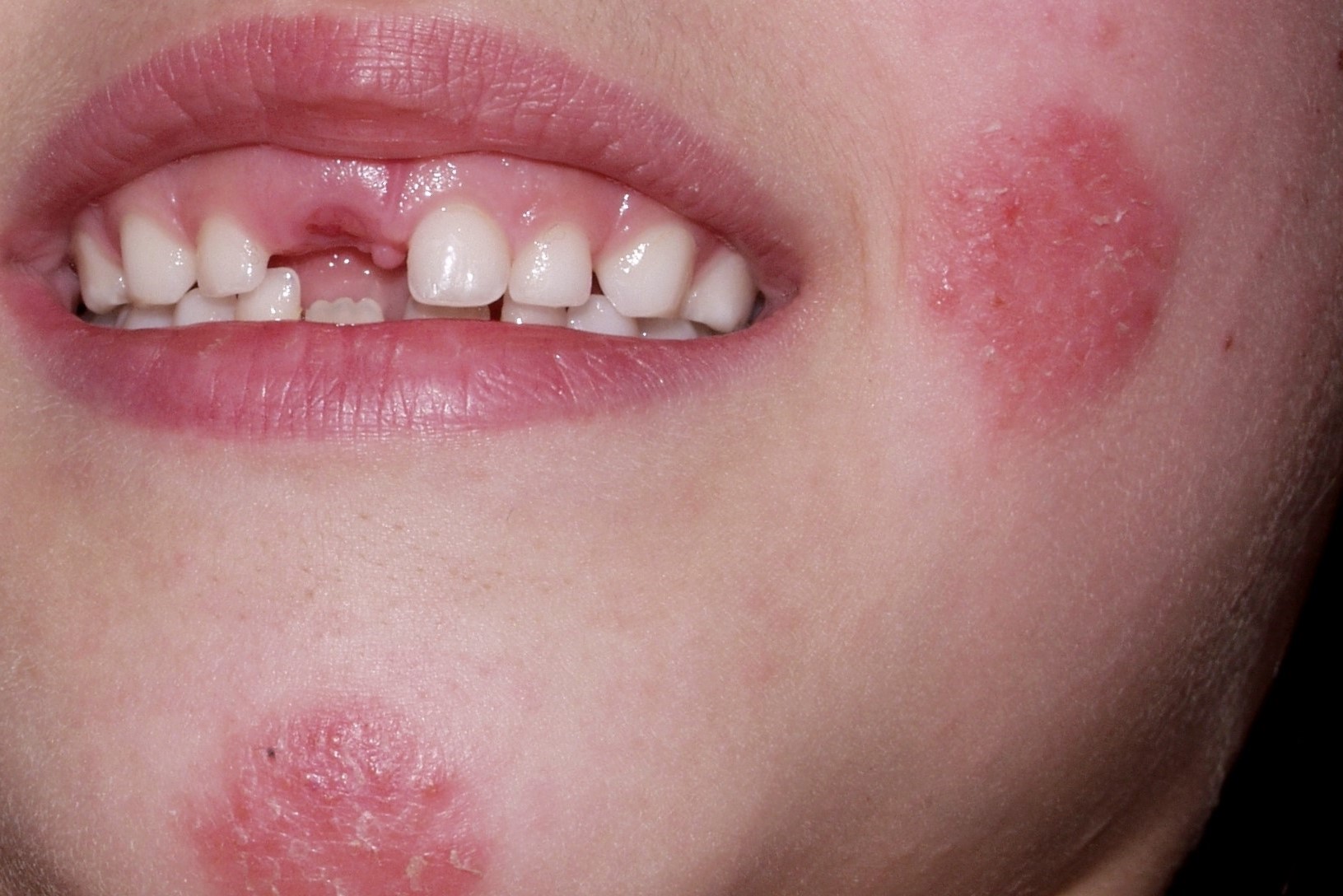
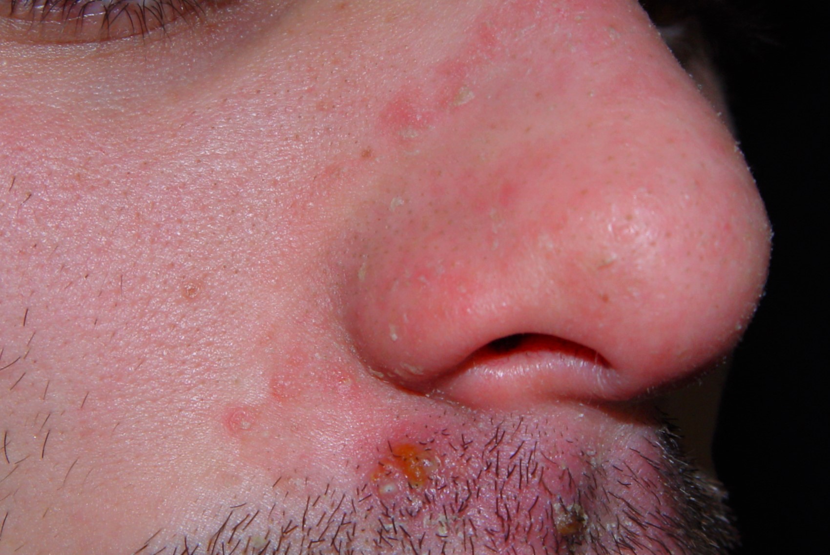
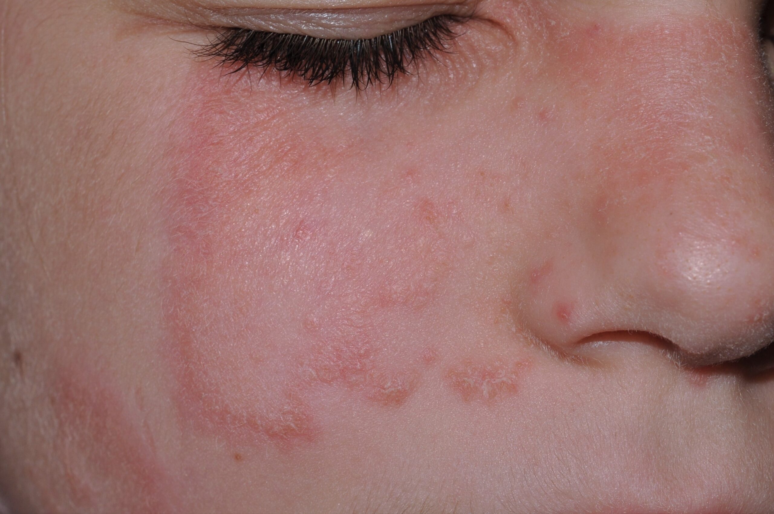
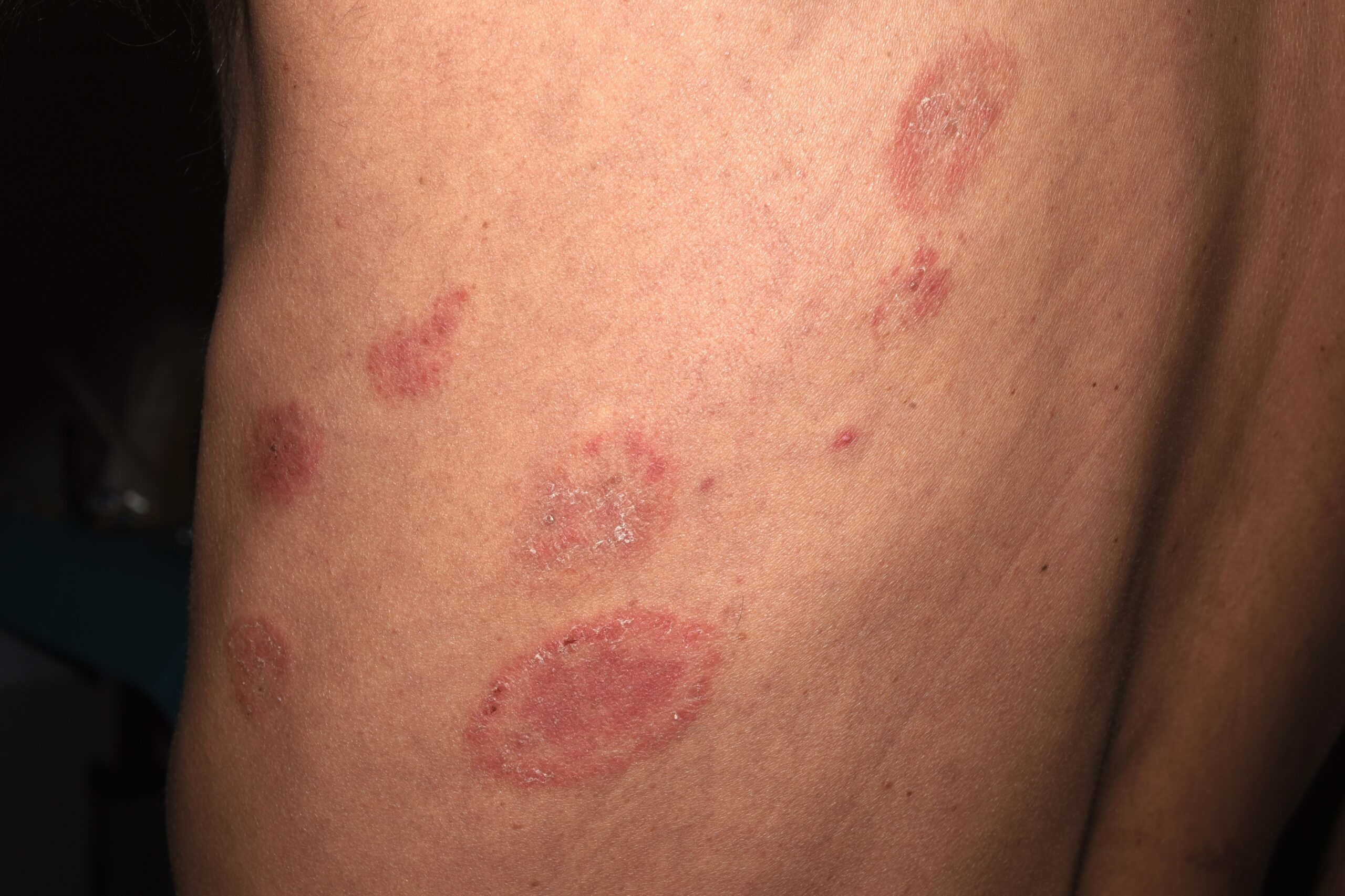
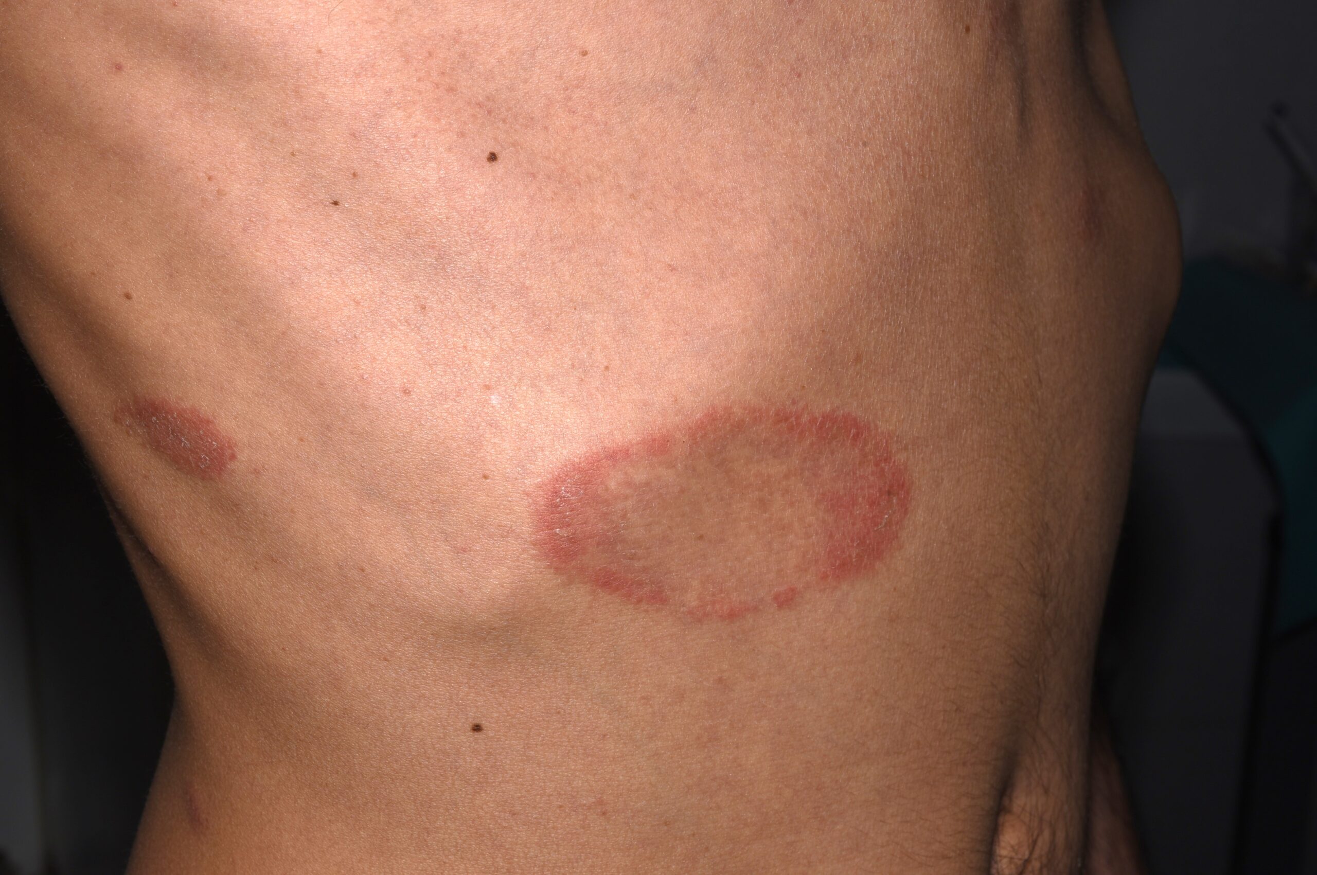
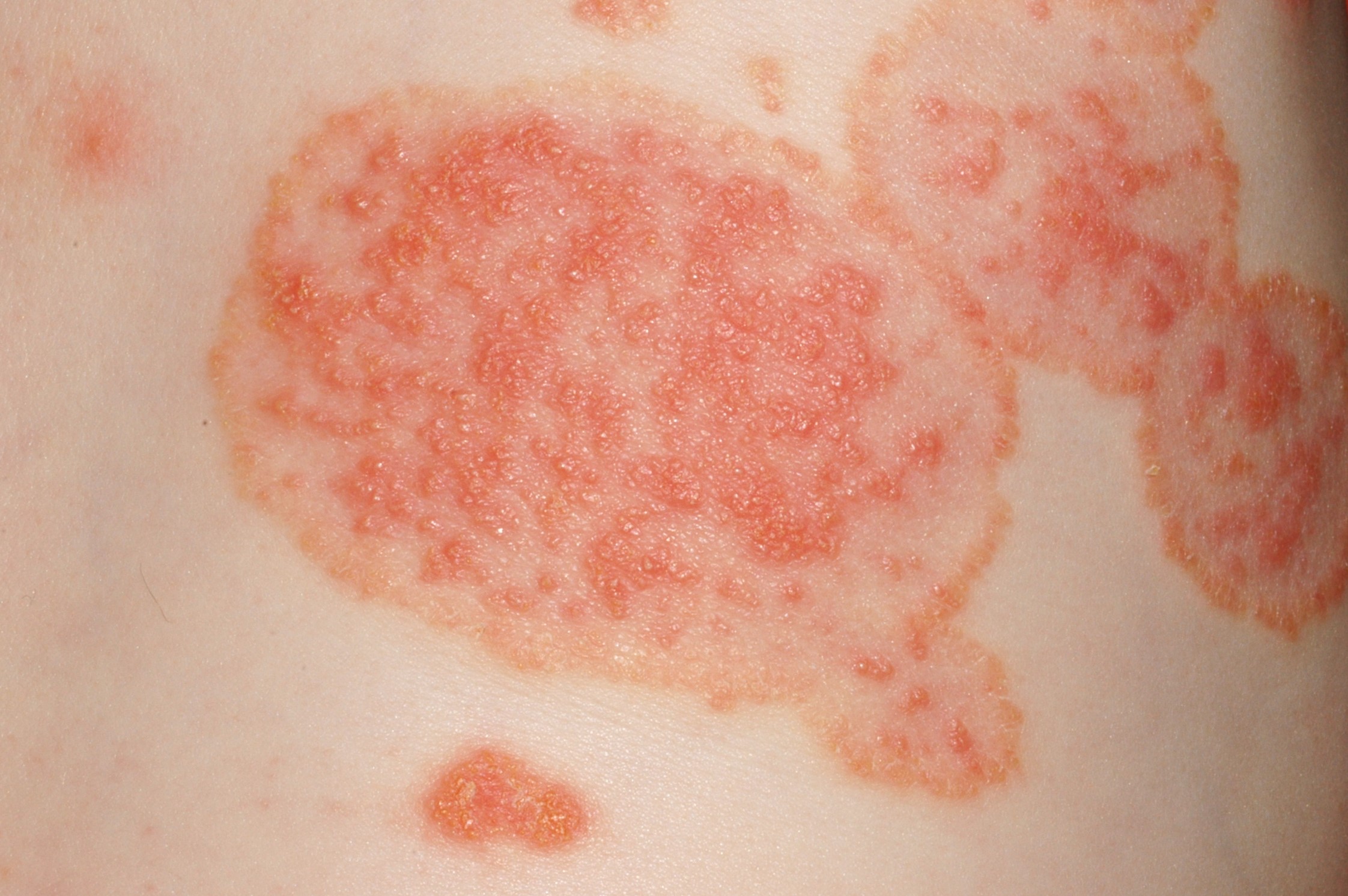
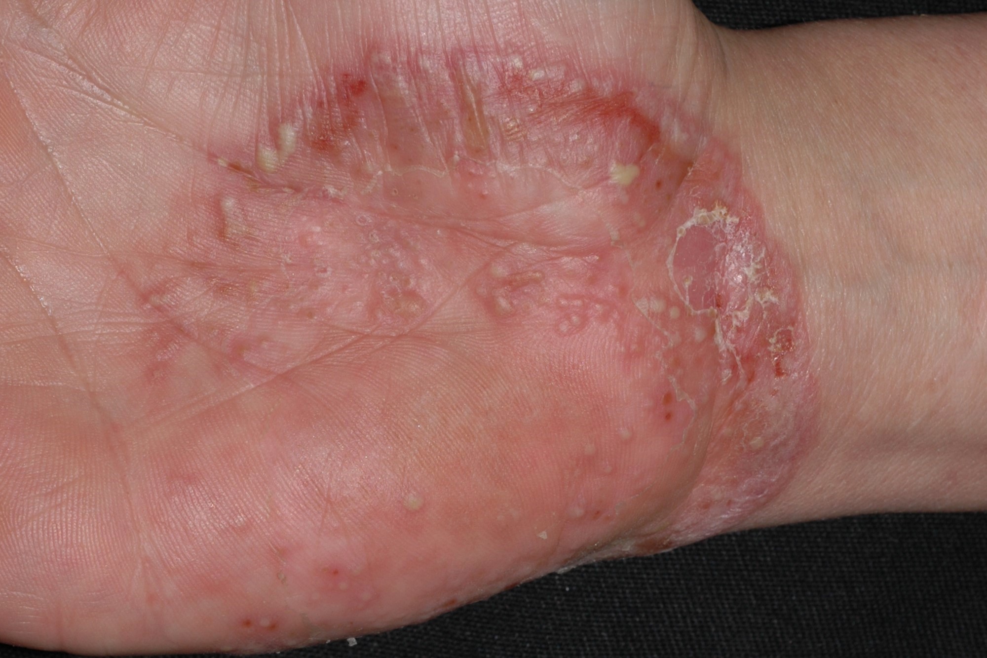
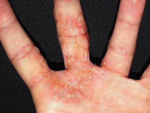
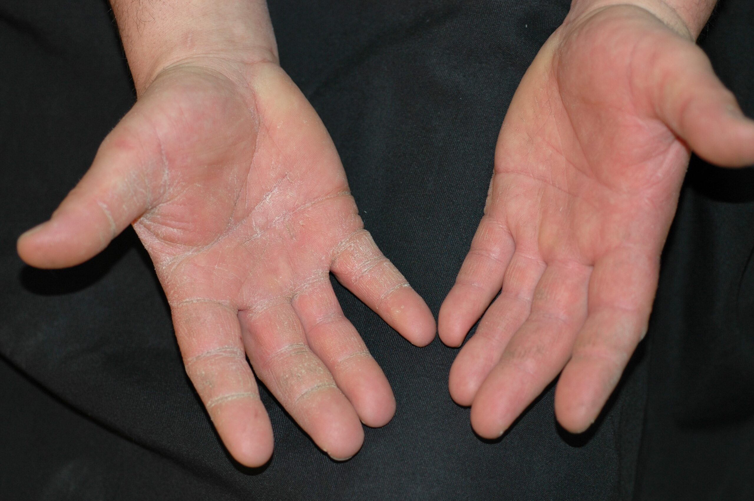
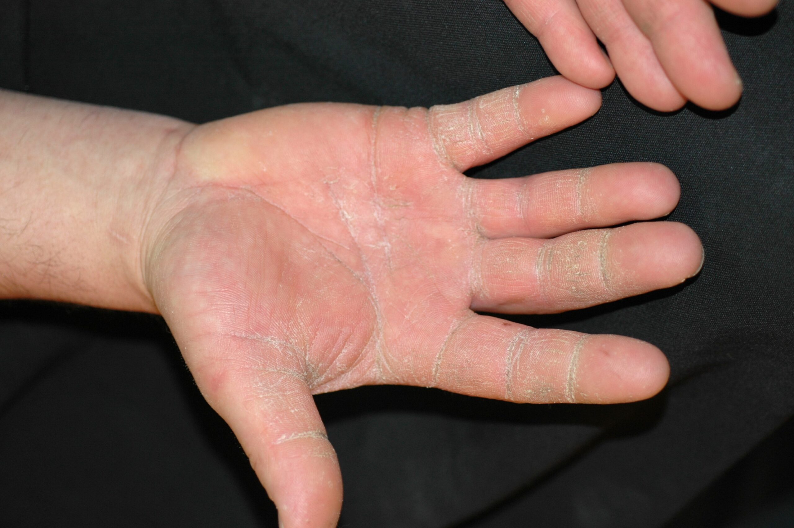
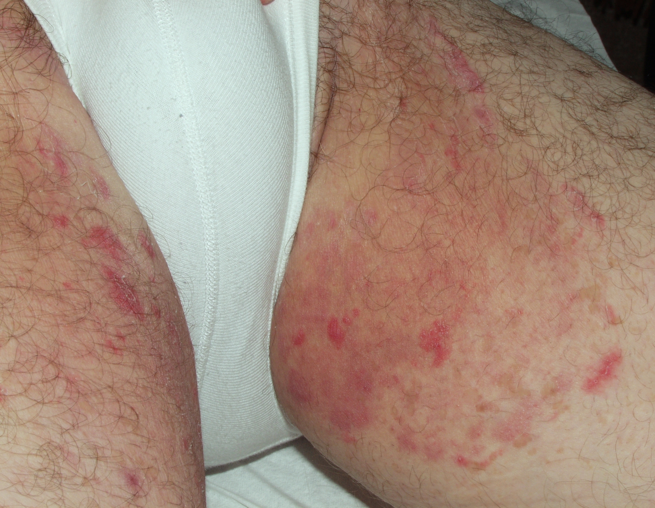
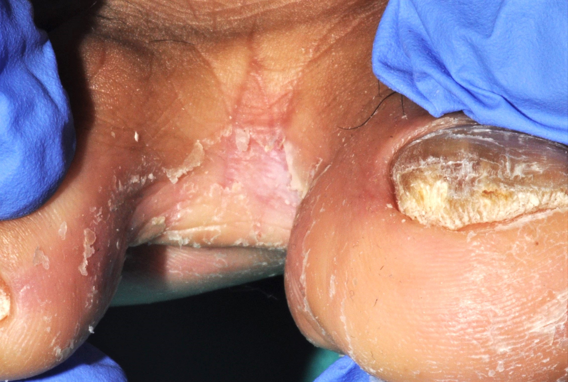
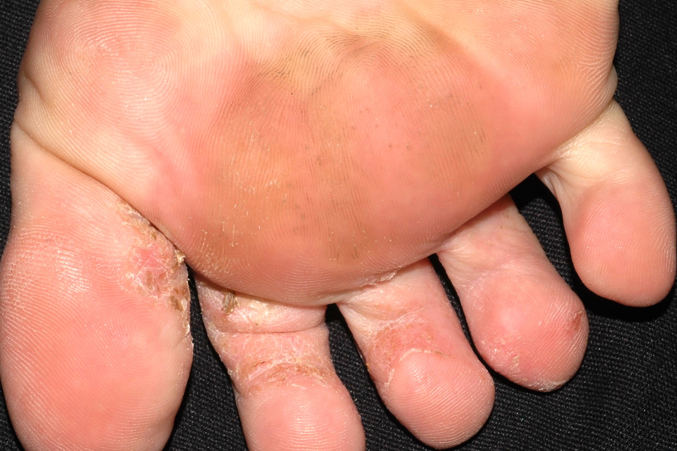
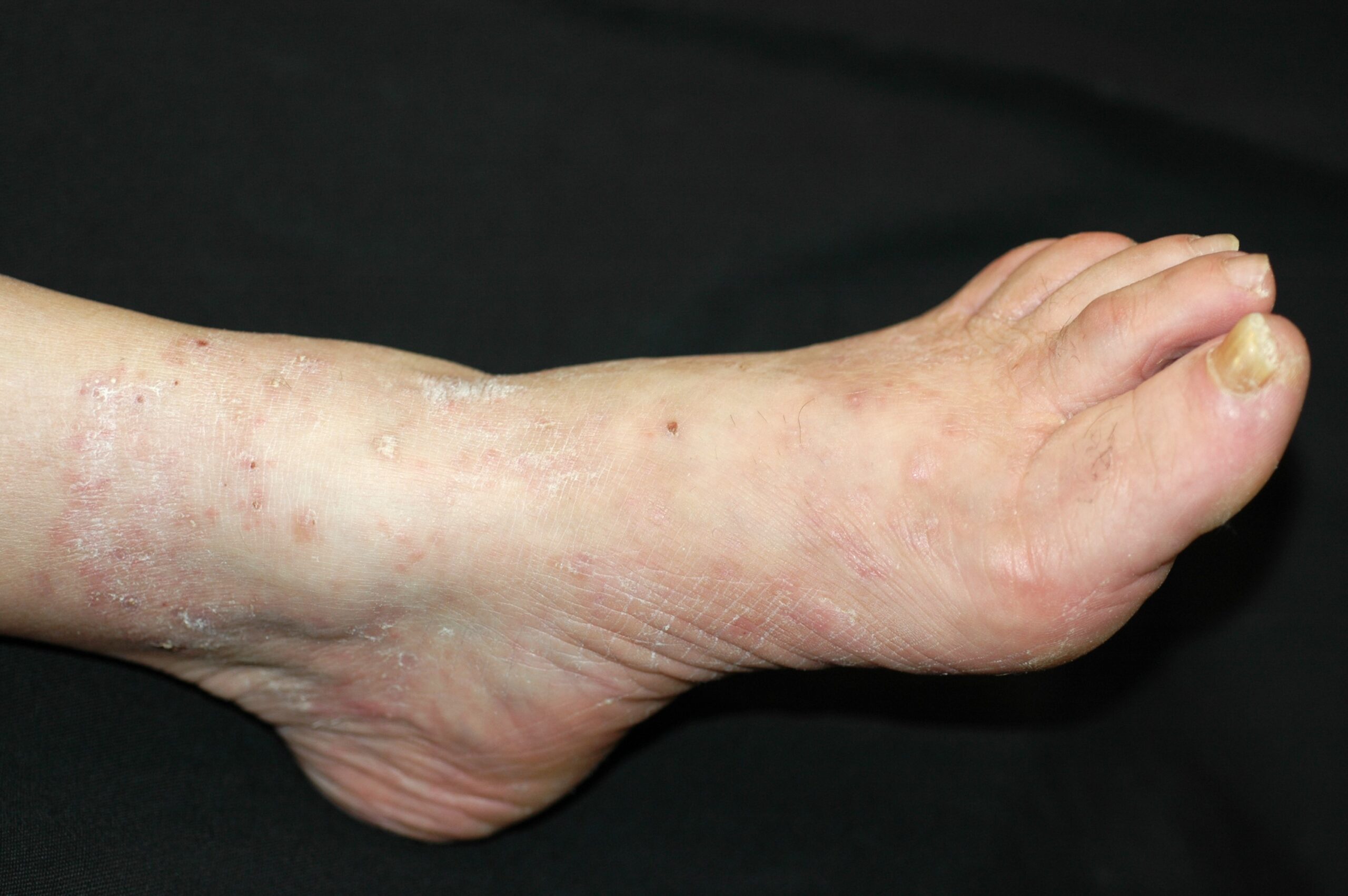
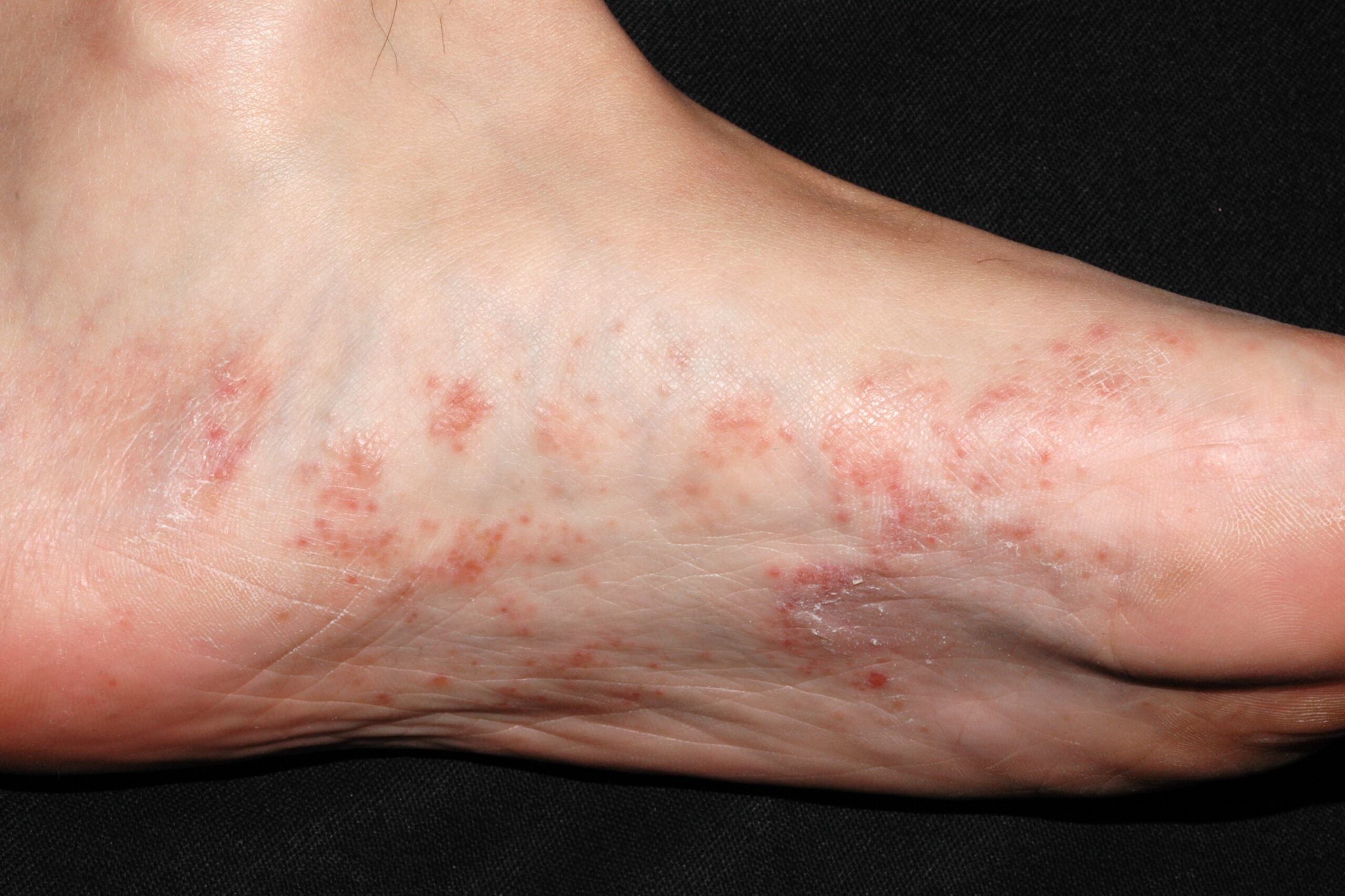
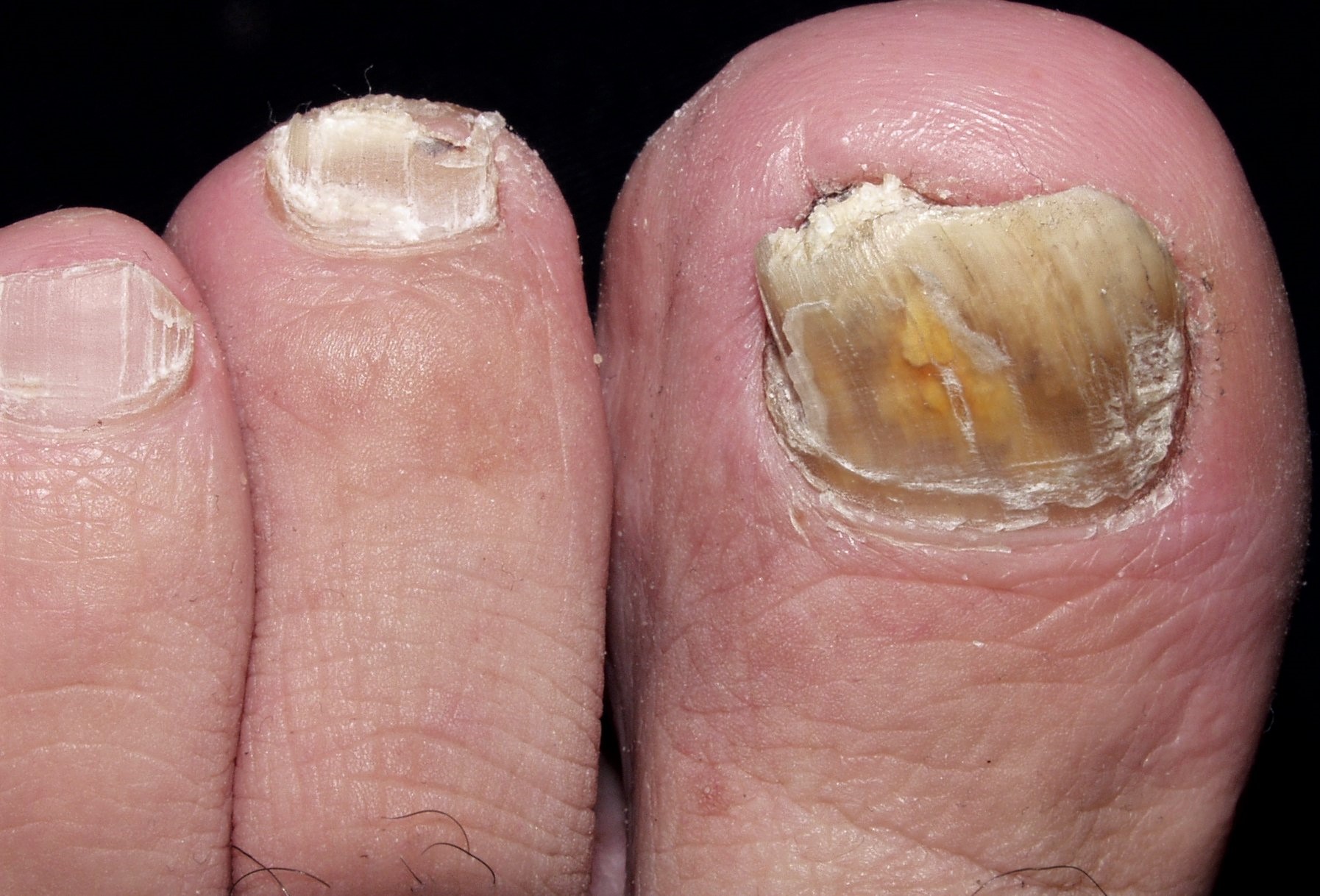
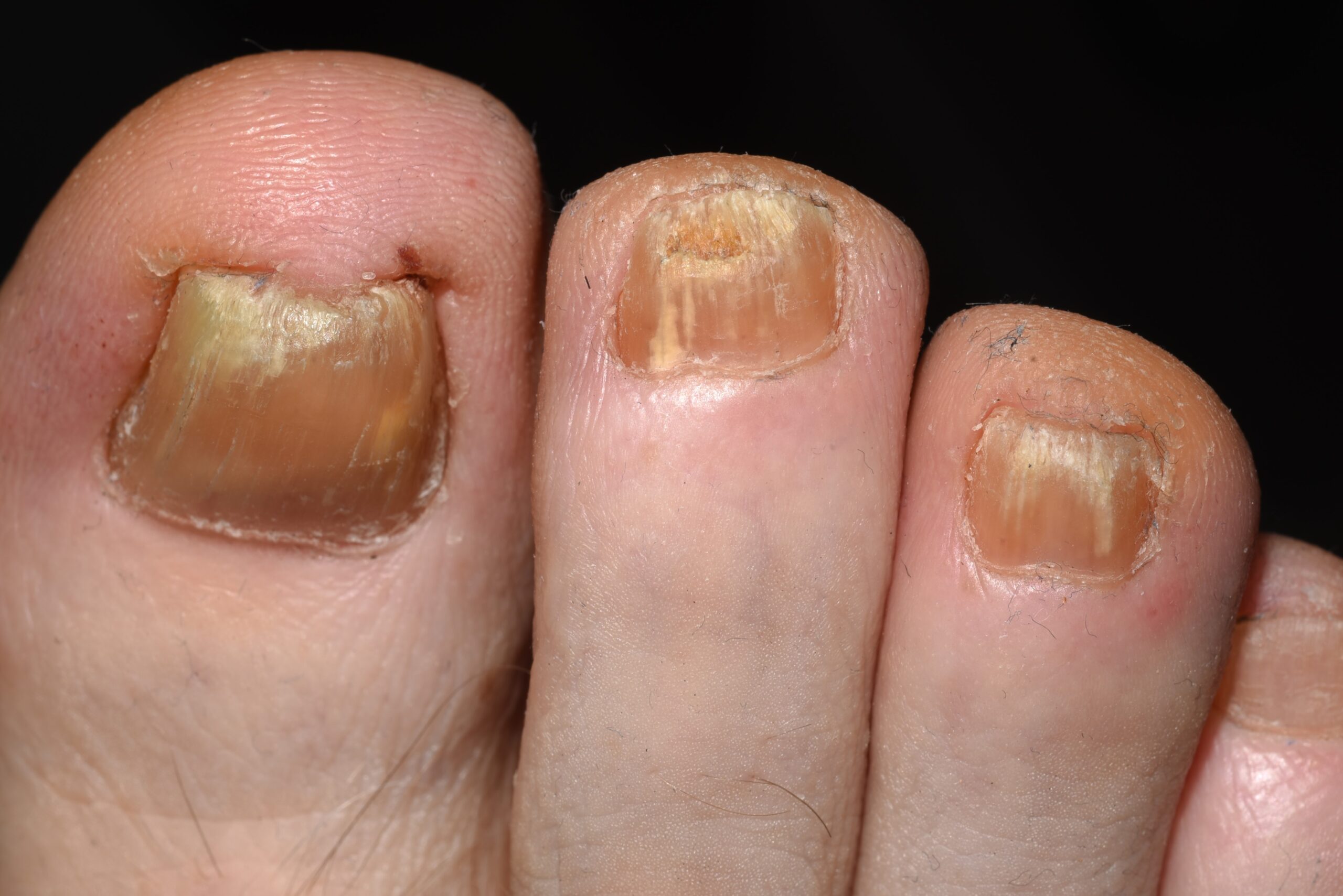
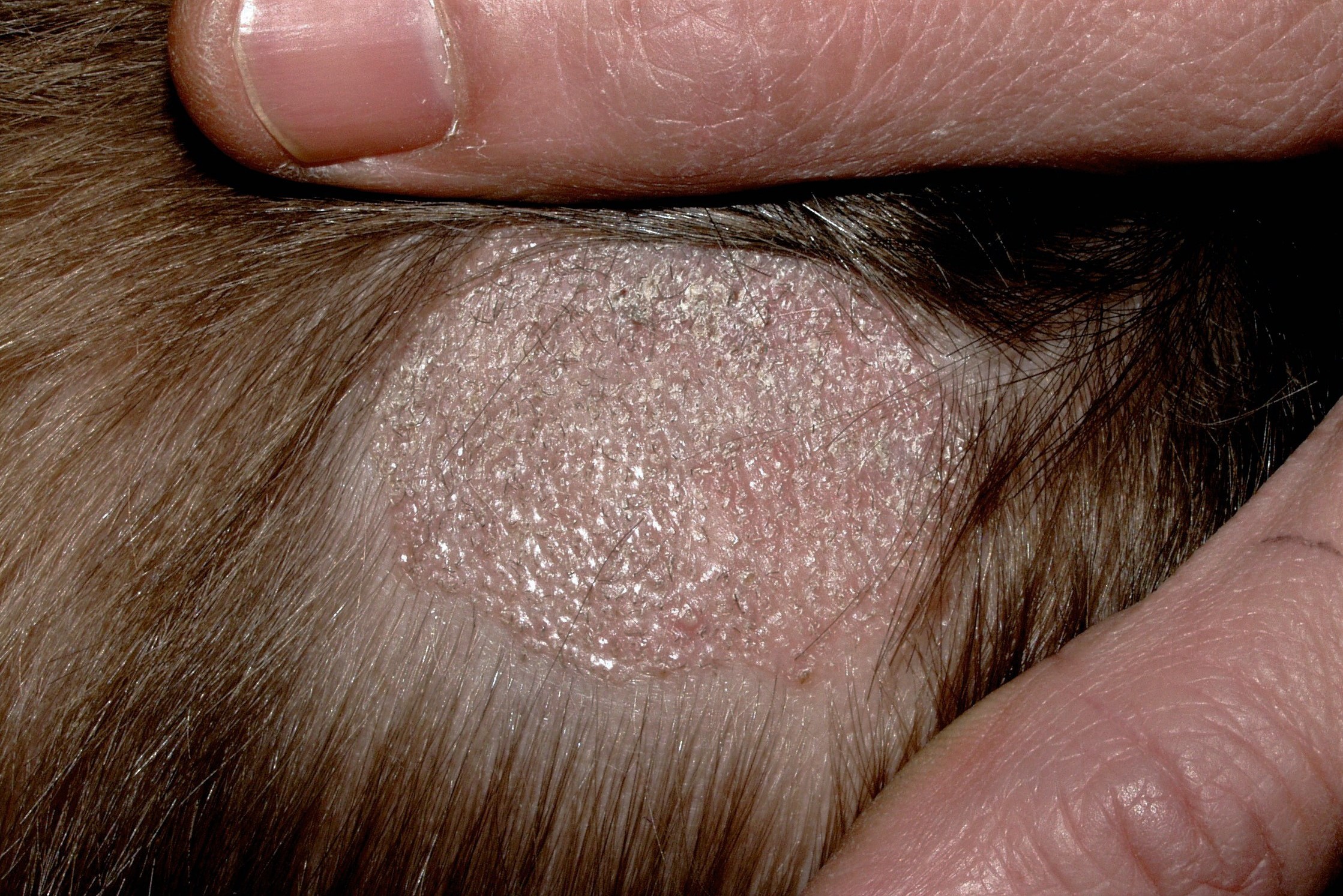
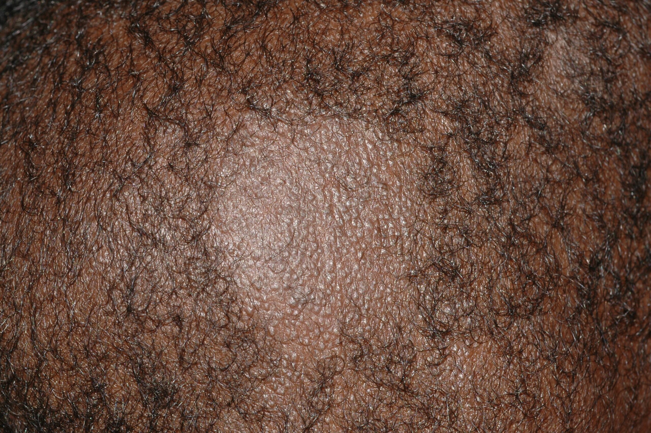
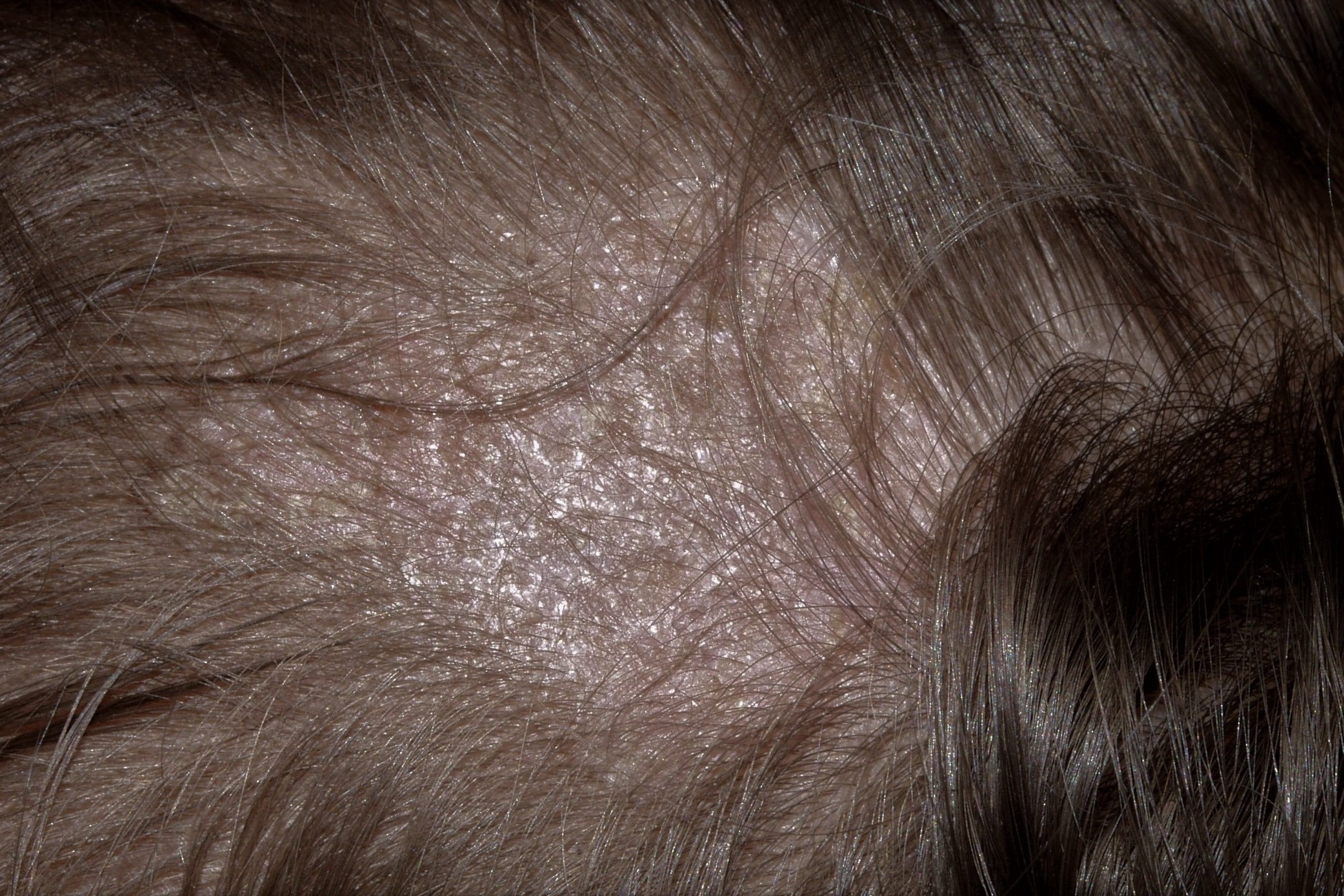
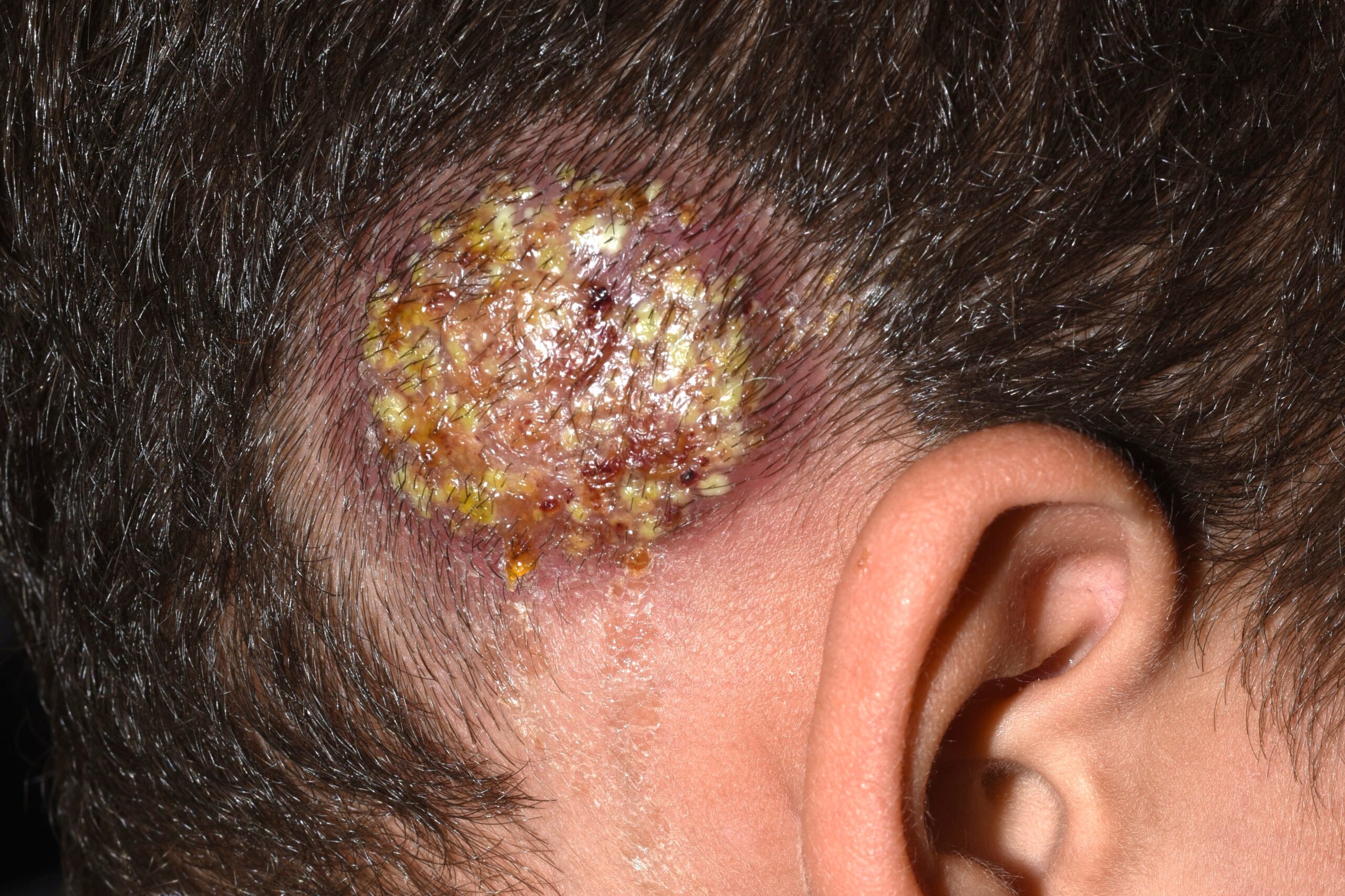
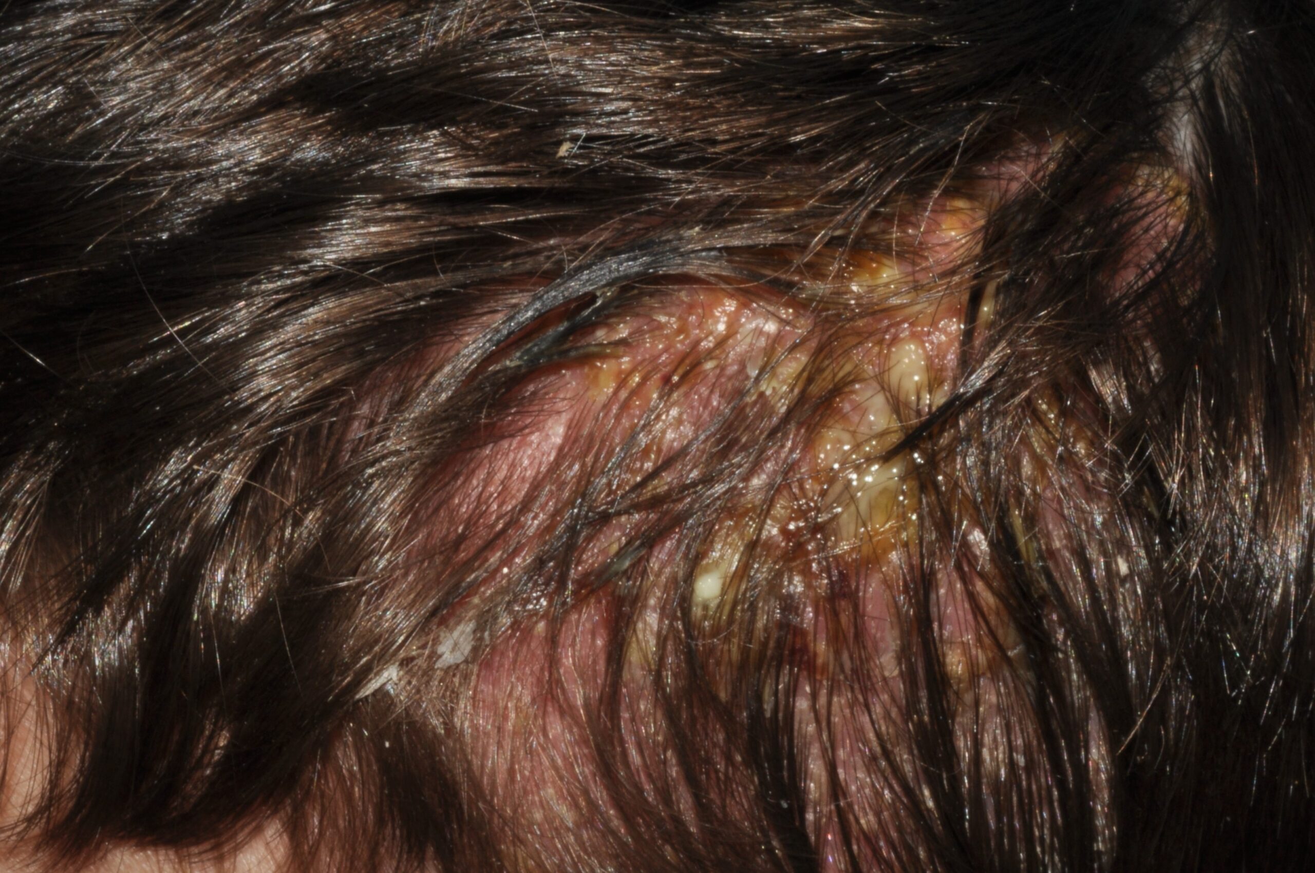
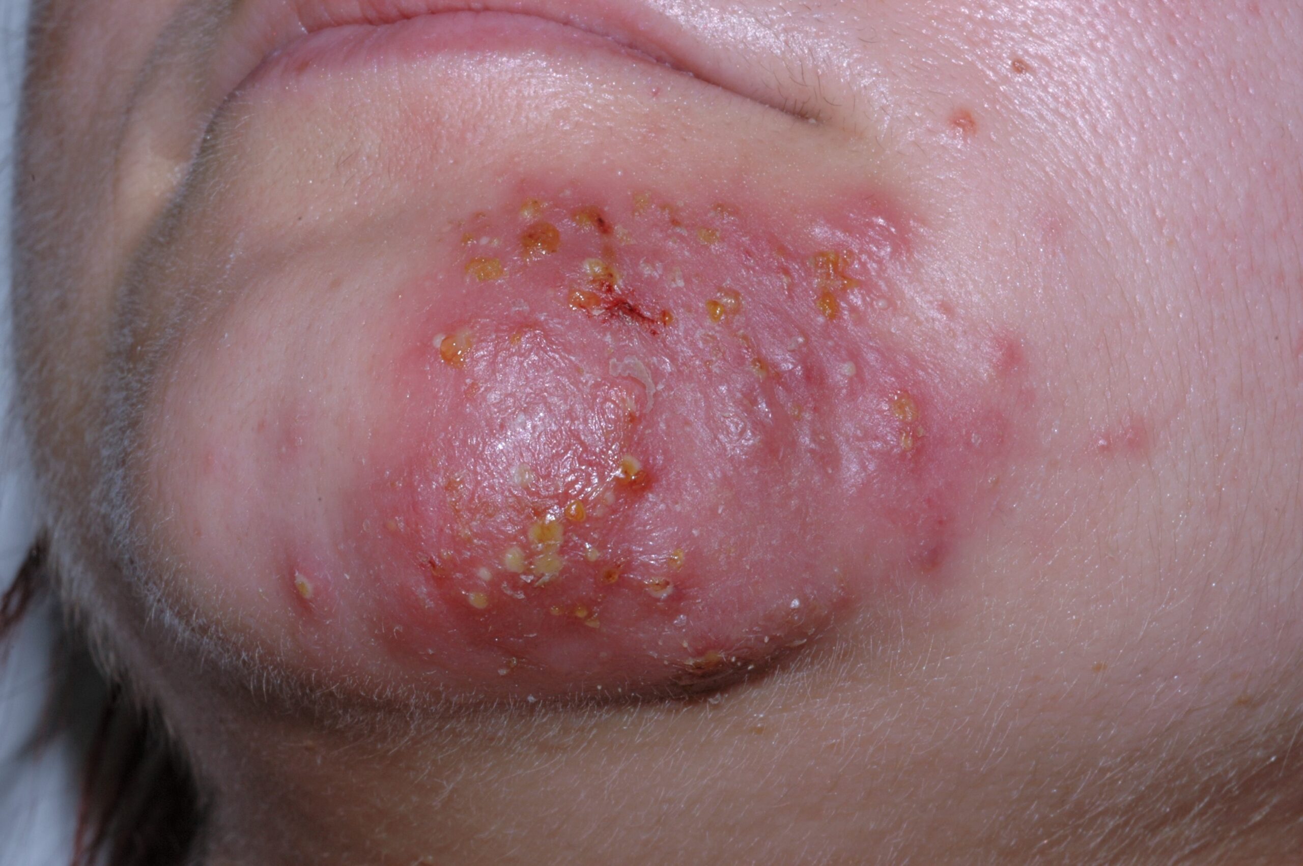
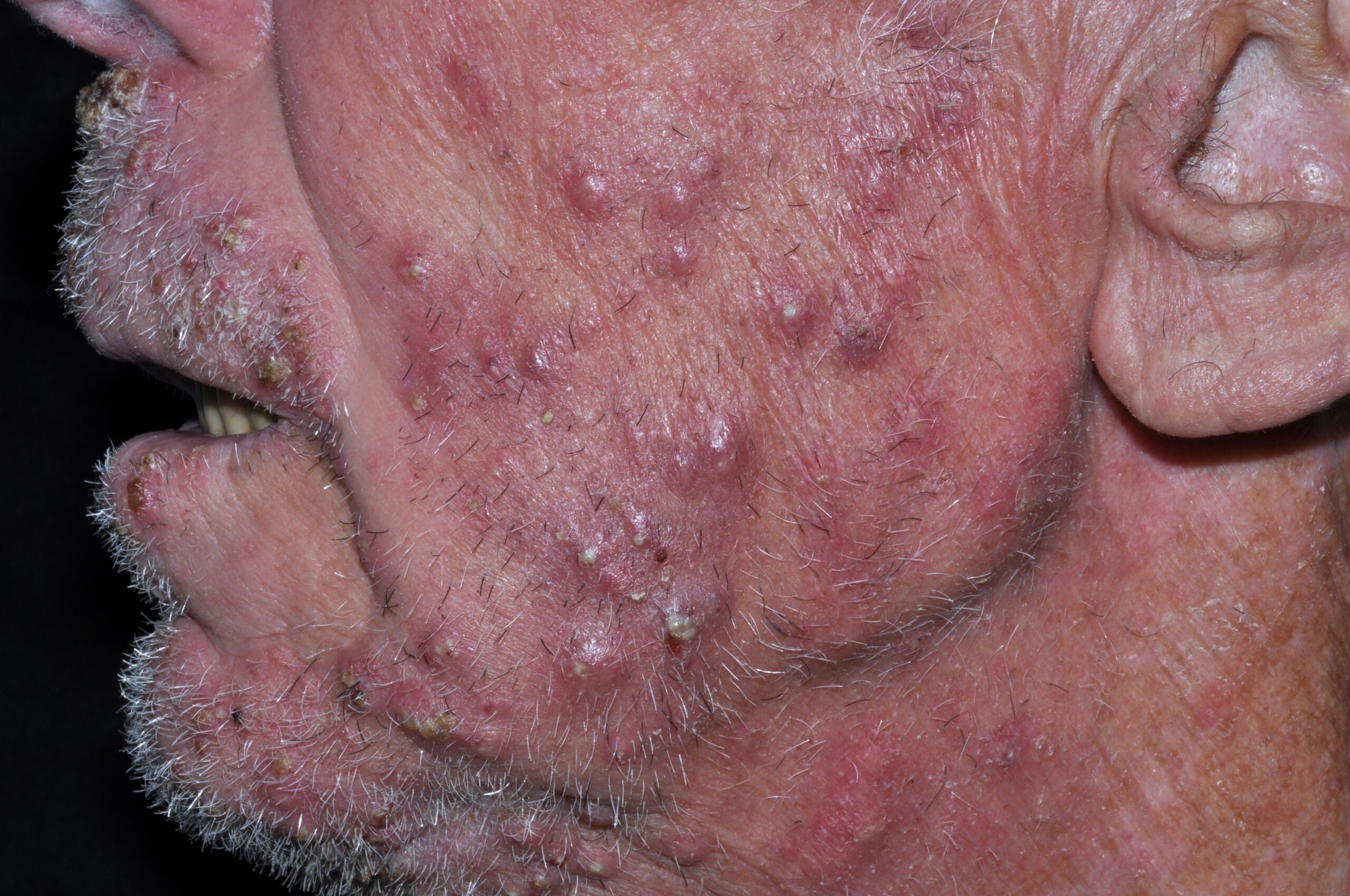
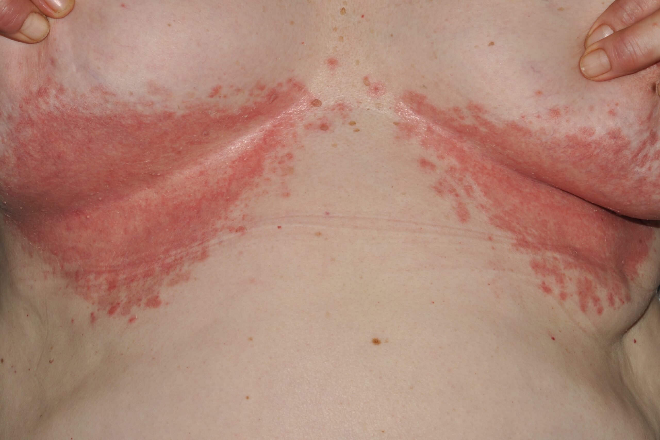
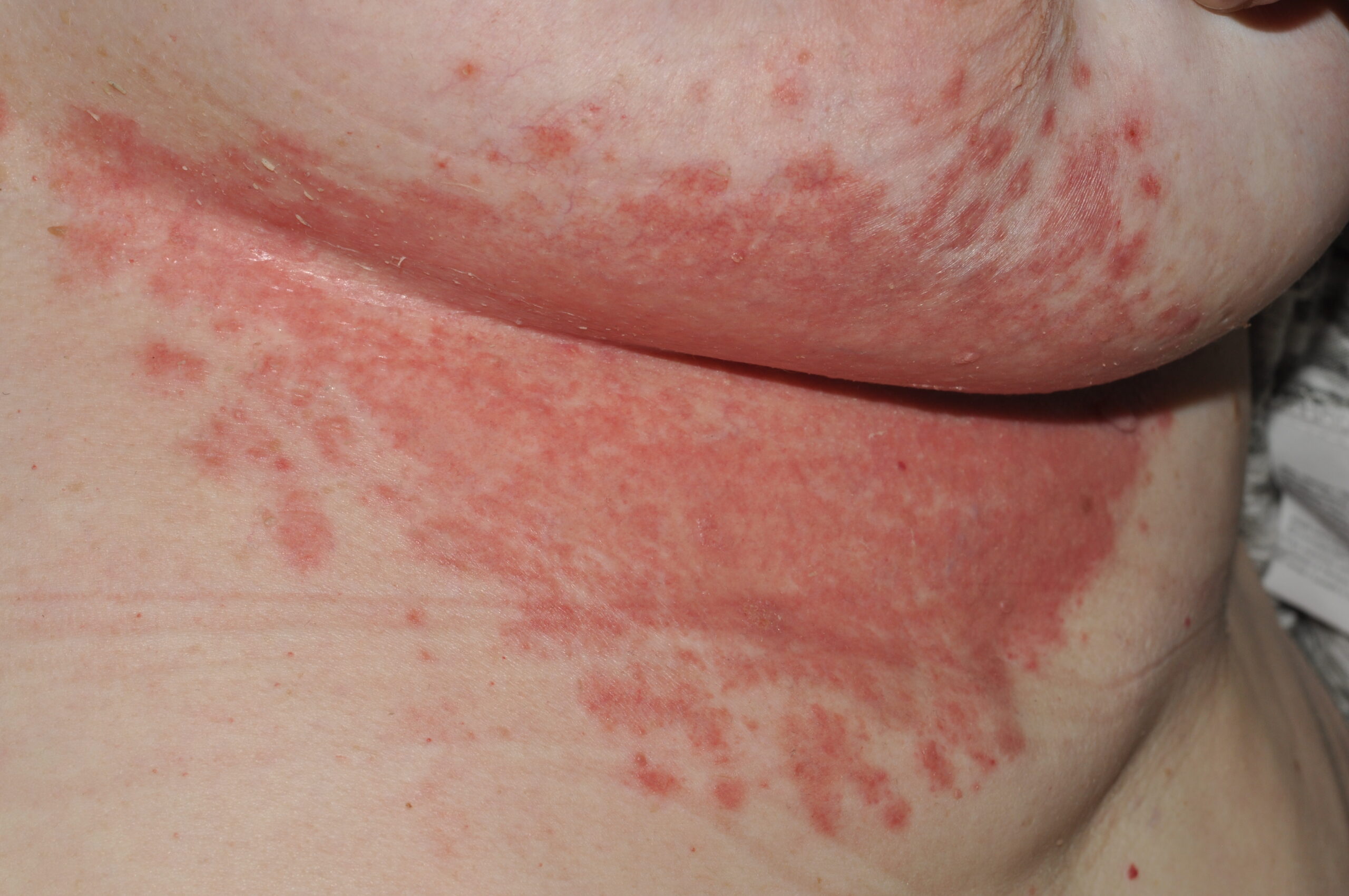
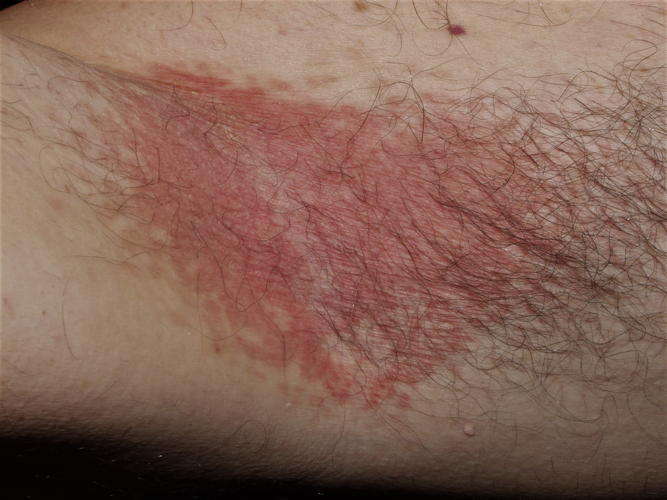
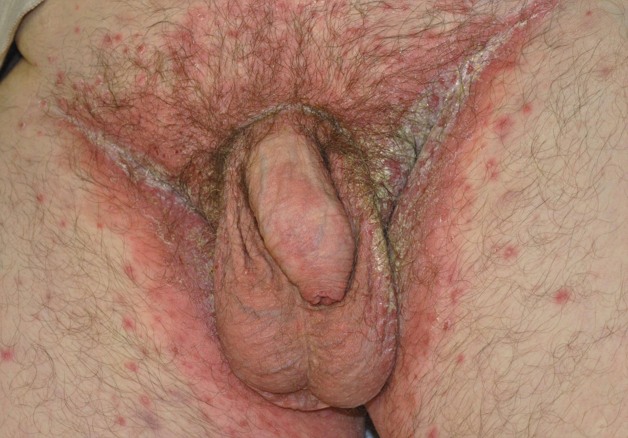
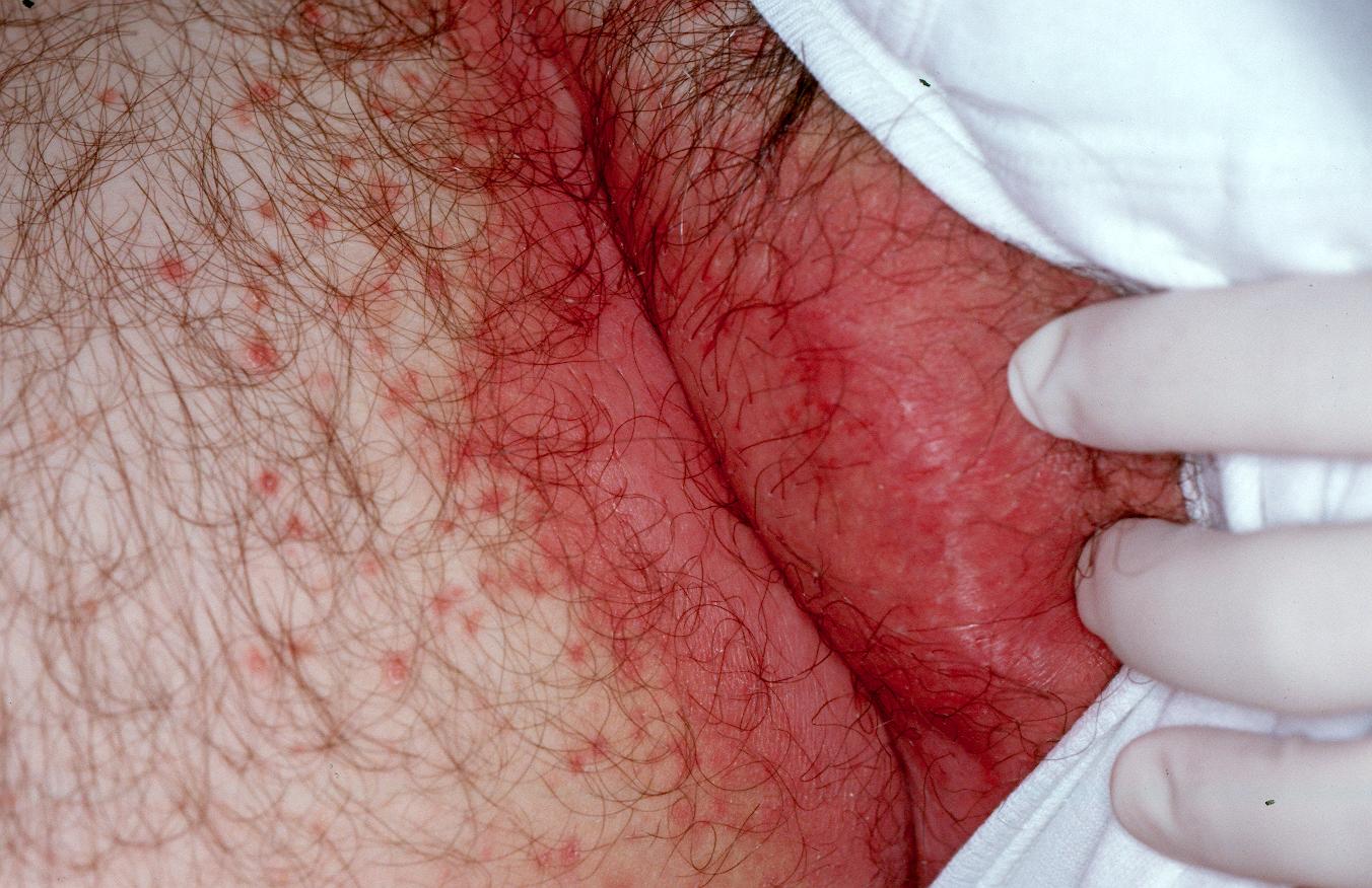
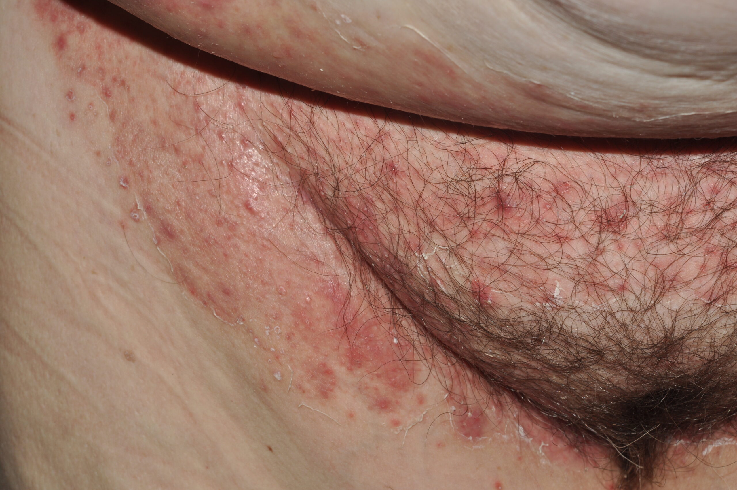
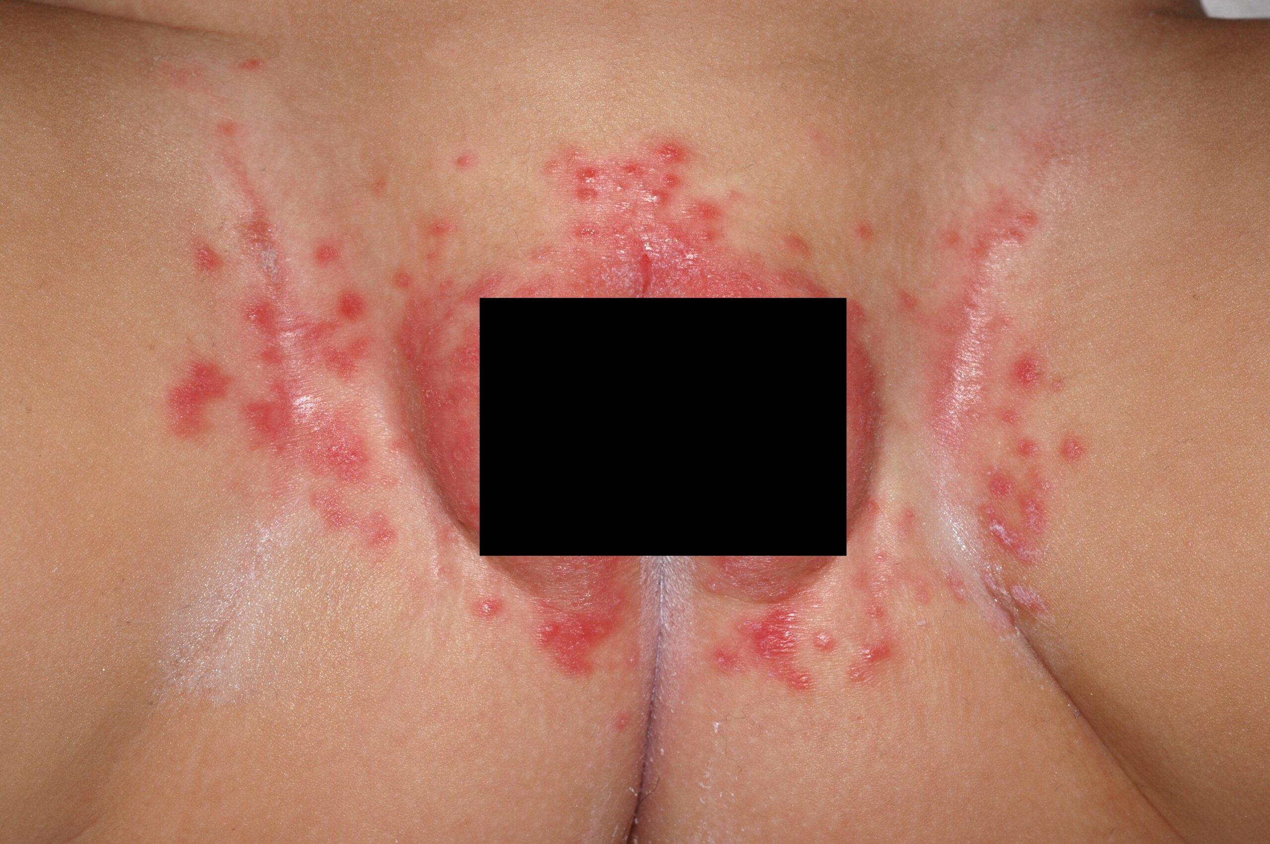
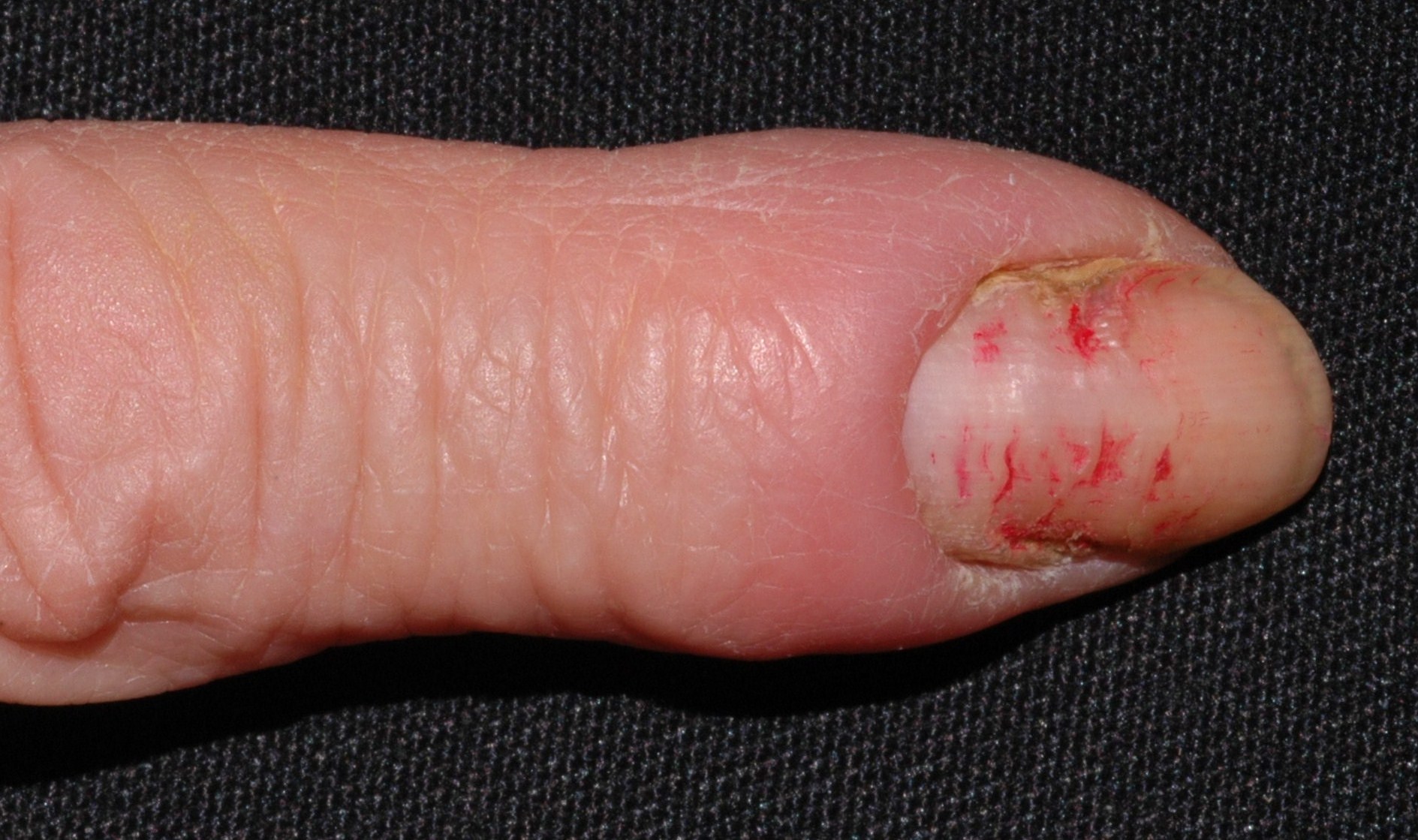
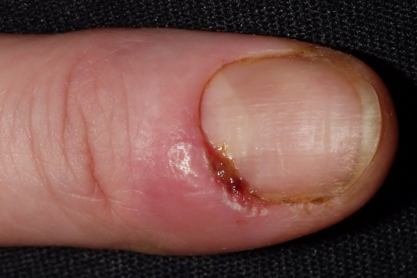
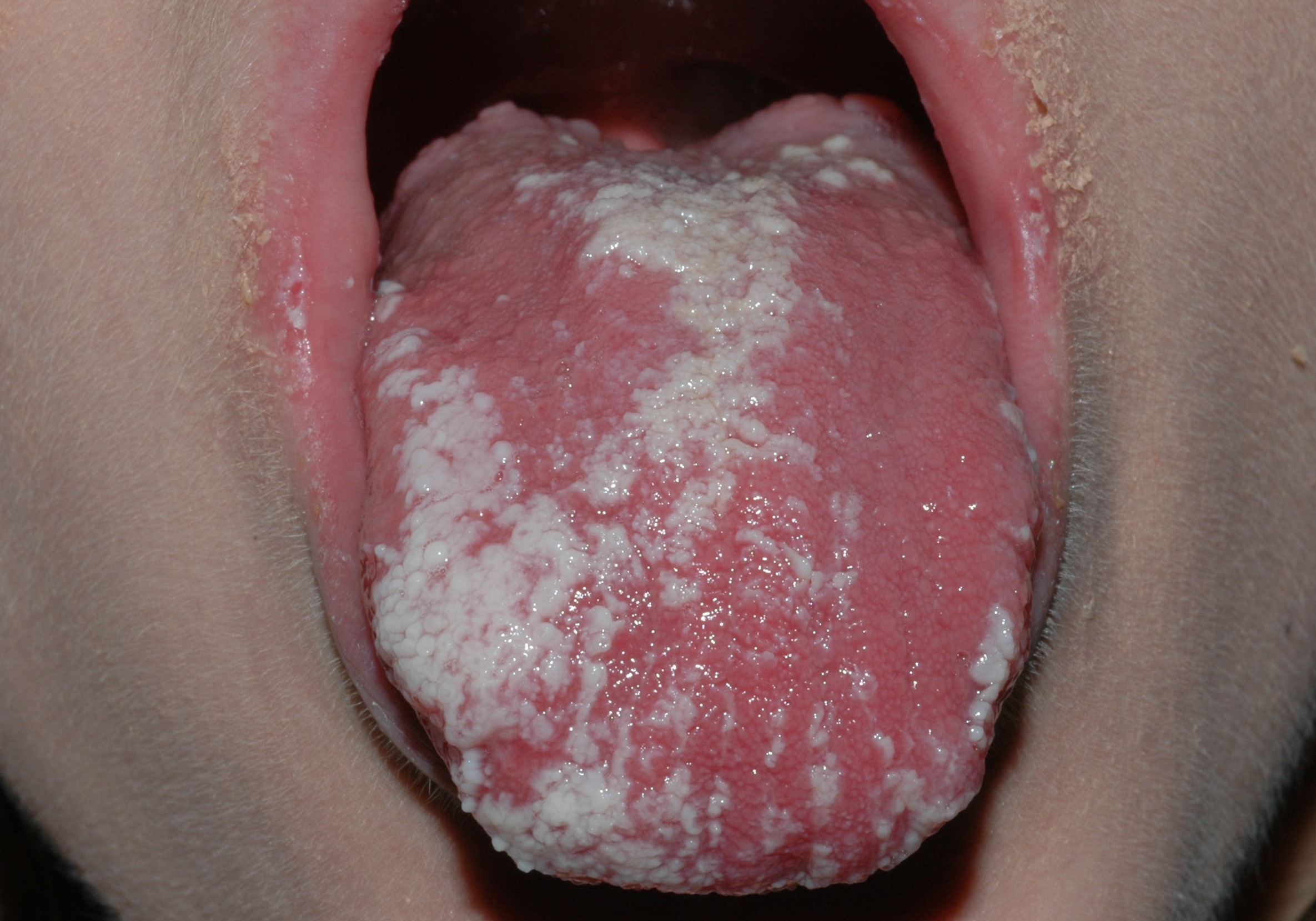
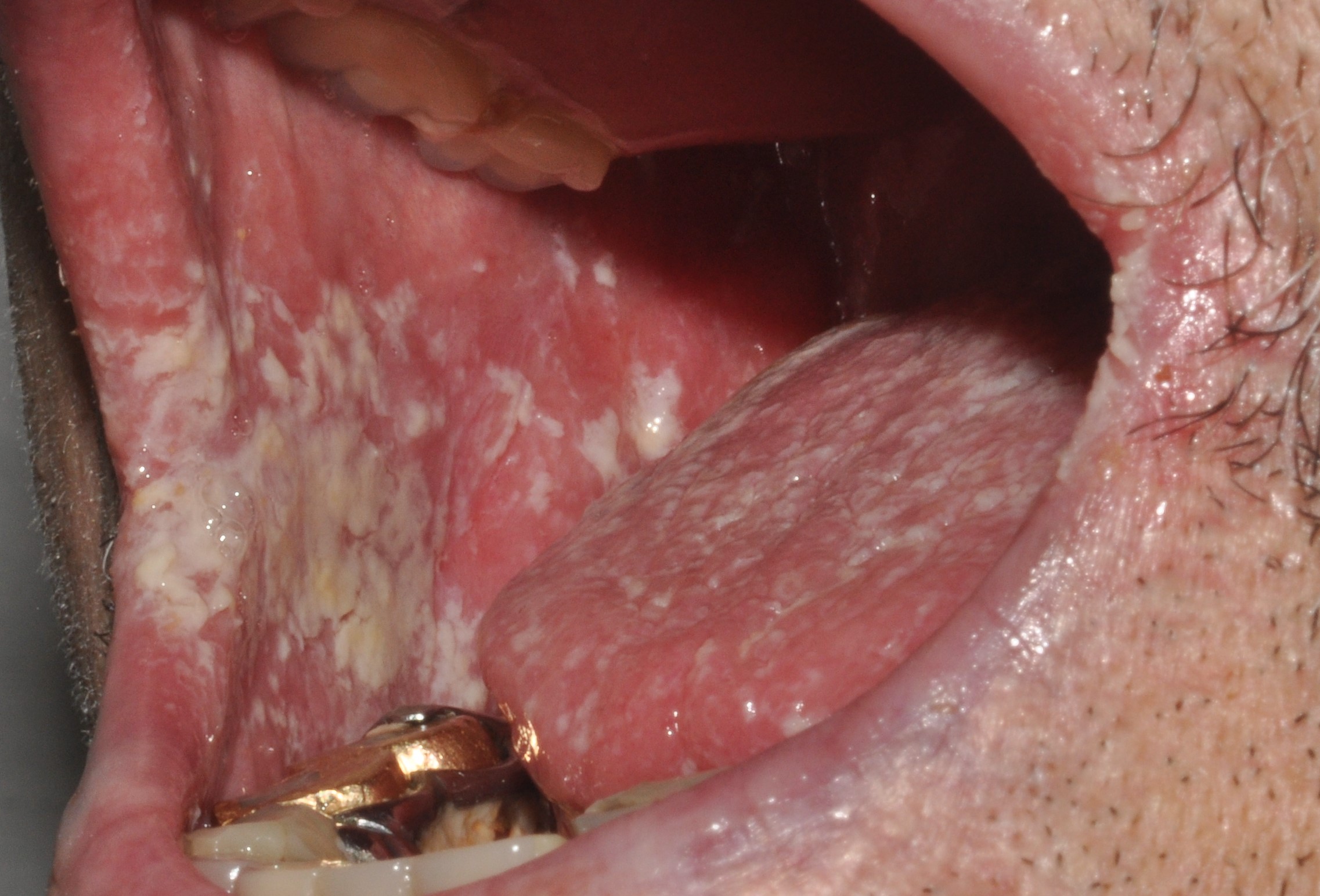
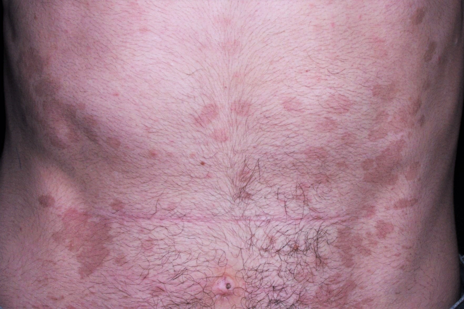
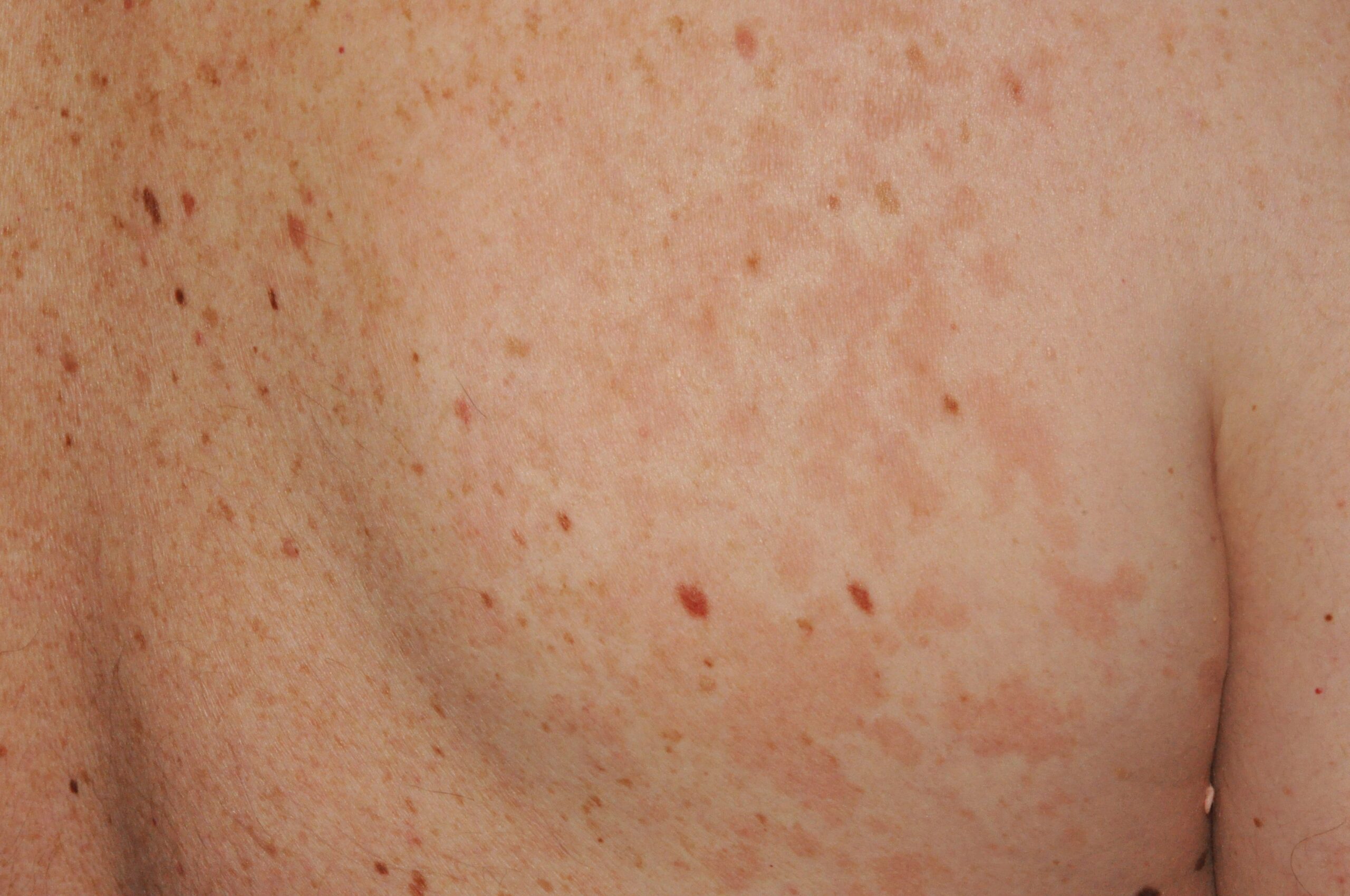
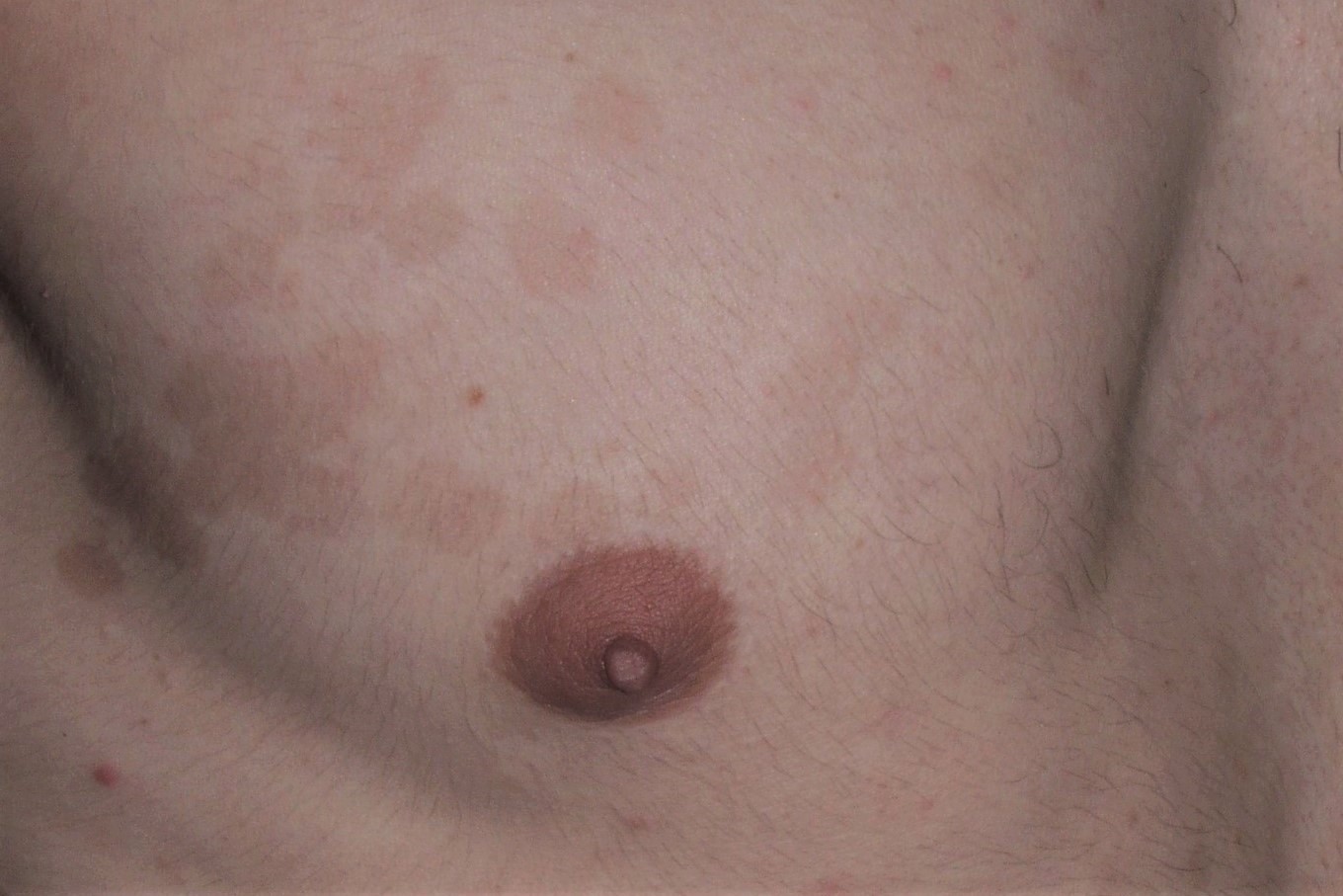
5. Biliografía
-Mora C, Tittensor DP, Adl S, Simpson AG, Worm B. How many species are there on Earth and in the ocean? PLoS Biol. 2011; 9(8):e1001127.
-Hawksworth DL, Lücking R. Fungal Diversity Revisited: 2.2 to 3.8 Million Species. Microbiol Spectr. 2017; 5(4). doi: 10.1128.
-Strickland AB, Shi M. Mechanisms of fungal dissemination. Cell Mol Life Sci. 2021; 78(7):3219-32.
-Yang J, Zhang KQ. Chitin Synthesis and Degradation in Fungi: Biology and Enzymes. Adv Exp Med Biol. 2019; 1142:153-167.
-Ahmad F, Yang G, Zhu Y, Poulsen M, Li W, Yu T, Mo J. Tripartite Symbiotic Digestion of Lignocellulose in the Digestive System of a Fungus-Growing Termite. Microbiol Spectr. 2022; 10(6):e0123422.
-Riquelme M, Aguirre J, Bartnicki-García S, Braus GH, Feldbrügge M, Fleig U, Hansberg W, Herrera-Estrella A, Kämper J, Kück U, Mouriño-Pérez RR, Takeshita N, Fischer R. Fungal Morphogenesis, from the Polarized Growth of Hyphae to Complex Reproduction and Infection Structures. Microbiol Mol Biol Rev. 2018; 82(2):e00068-17.
-Sudbery PE. Growth of Candida albicans hyphae. Nat Rev Microbiol. 2011; 9(10):737-48.
-Kumamoto CA, Vinces MD. Contributions of hyphae and hypha-co-regulated genes to Candida albicans virulence. Cell Microbiol. 2005; 7(11):1546-54.
-Li J, Feng Y, Liu C, Yang Z, de Hoog S, Qu Y, Chen B, Li D, Xiong H, Shi D. Presence of Malassezia Hyphae Is Correlated with Pathogenesis of Seborrheic Dermatitis. Microbiol Spectr. 2022; 10(1):e0116921.
-Martinez-Rossi NM, Peres NTA, Bitencourt TA, Martins MP, Rossi A. State-of-the-Art Dermatophyte Infections: Epidemiology Aspects, Pathophysiology, and Resistance Mechanisms. J Fungi (Basel). 2021; 7(8):629.
-Rokas A. Evolution of the human pathogenic lifestyle in fungi. Nat Microbiol. 2022; 7(5):607-619.
-Brown GD, Ballou ER, Bates S, Bignell EM, Borman AM, Brand AC, Brown AJP, Coelho C, Cook PC, Farrer RA, Govender NP, Gow NAR, Hope W, Hoving JC, Dangarembizi R, Harrison TS, Johnson EM, Mukaremera L, Ramsdale M, Thornton CR, Usher J, Warris A, Wilson D. The pathobiology of human fungal infections. Nat Rev Microbiol. 2024; 22(11):687-704.
-Qin Q, Su J, Liu J, Chen R, Wei W, Yuan Z, Lai S, Duan R, Lai J, Ye L, Liang H, Jiang J. Global, regional, and national burden of fungal skin diseases in 204 countries and territories from 1990 to 2021: An analysis of the global burden of disease study 2021. Mycoses. 2024; 67(8):e13787
-Sei Y. 2011 Epidemiological Survey of Dermatomycoses in Japan. Med Mycol J. 2015; 56:J129-35.
-Sharma B, Nonzom S. Superficial mycoses, a matter of concern: Global and Indian scenario-an updated analysis. Mycoses. 2021; 64(8):890-908.
-Hawkins DM, Smidt AC. Superficial fungal infections in children. Pediatr Clin North Am. 2014; 61(2):443-55.
-Nowicka D, Nawrot U. Tinea pedis-An embarrassing problem for health and beauty-A narrative review. Mycoses. 2021; 64(10):1140-1150.
-Barac A, Stjepanovic M, Krajisnik S, Stevanovic G, Paglietti B, Milosevic B. Dermatophytes: Update on Clinical Epidemiology and Treatment. Mycopathologia. 2024; 189(6):101.
-Celestrino GA, Verrinder Veasey J, Benard G, Sousa MGT. Host immune responses in dermatophytes infection. Mycoses. 2021; 64(5):477-483.
-de Hoog GS, Dukik K, Monod M, Packeu A, Stubbe D, Hendrickx M, Kupsch C, Stielow JB, Freeke J, Göker M, Rezaei-Matehkolaei A, Mirhendi H, Gräser Y. Toward a novel multilocus phylogenetic taxonomy for the dermatophytes. Mycopathologia 2017, 182, 5-31.
-Balaguer-Franch I, Hernández de la Torre-Ruiz E, Baniandrés-Rodríguez O. Generalized dermatophytosis and tinea capitis mimicking psoriasis in a patient with long-term infliximab treatment. Clin Exp Dermatol. 2021; 46(4):750-751.
-Si H, Li Y, Huang Z, Cui Y, Li S. Erythroderma combined with deeper dermal dermatophytosis due to Trichophyton rubrum in a patient with myasthenia gravis: first case report and literature review. BMC Infect Dis. 2023; 23(1):789.
-Dellière S, Joannard B, Benderdouche M, Mingui A, Gits-Muselli M, Hamane S, Alanio A, Petit A, Gabison G, Bagot M, Bretagne S. Emergence of Difficult-to-Treat Tinea Corporis Caused by Trichophyton mentagrophytes Complex Isolates, Paris, France. Emerg Infect Dis. 2022; 28(1):224-228.
-Jabet A, Brun S, Normand AC, Imbert S, Akhoundi M, Dannaoui E, Audiffred L, Chasset F, Izri A, Laroche L, Piarroux R, Bachmeyer C, Hennequin C, Sabater AM. Extensive Dermatophytosis Caused by Terbinafine-Resistant Trichophyton indotineae, France. Emerg Infect Dis. 2022;28(1):229-233.
-Jabet A, Normand AC, Brun S, Dannaoui E, Bachmeyer C, Piarroux R, Hennequin C, Moreno-Sabater A. Trichophyton indotineae, from epidemiology to therapeutic. J Mycol Med. 2023; 33(3):101383.
-Boehm TMSA, Mueller RS. Dermatophytosis in dogs and cats – an update. Tierarztl Prax Ausg K Kleintiere Heimtiere. 2019; 47(4):257-268.
-Monod M, Fratti M, Mignon B, Baudraz-Rosselet F. [Dermatophytes transmitted by pets and cattle]. Rev Med Suisse. 2014; 10(424):749-53.
-Uhrlaß S, Mayser P, Koch D, Mütze H, Krüger C, Schulze I, Nenoff P. Zoophile Dermatophyten während der Corona-Pandemie in Deutschland. Dermatologie (Heidelb). 2023; 74(6):430-439.
-Long S, Carveth H, Chang YM, O’Neill D, Bond R. Isolation of dermatophytes from dogs and cats in the South of England between 1991 and 2017. Vet Rec. 2020; 187(10):e87
-Lanternier F, Pathan S, Vincent QB, Liu L, Cypowyj S, Prando C, Migaud M, Taibi L, Ammar-Khodja A, Stambouli OB, Guellil B, Jacobs F, Goffard JC, Schepers K, Del Marmol V, Boussofara L, Denguezli M, Larif M, Bachelez H, Michel L, Lefranc G, Hay R, Jouvion G, Chretien F, Fraitag S, Bougnoux ME, Boudia M, Abel L, Lortholary O, Casanova JL, Picard C, Grimbacher B, Puel A. Deep dermatophytosis and inherited CARD9 deficiency. N Engl J Med. 2013; 369(18):1704-1714.
-Antuori A, Fernández G, Fernández A, Alcaide M, Boada A, Bielsa MI, Romaní N, Matas L. Epidemiology of dermatophytic infections between 2008 and 2017 in Barcelona, Spain. Enferm Infecc Microbiol Clin (Engl Ed). 2019; 37(10):642-647.
-Reichert-Pénétrat S, Contet-Audonneau N, Barbaud A, Schurra JP, Fortier B, Schmutz JL. Epidemiology of dermatophytoses in children living in northeast France: a 5-year study. Pediatr Dermatol. 2002; 19(2):103-5.
-Courtellemont L, Chevrier S, Degeilh B, Belaz S, Gangneux JP, Robert-Gangneux F. Epidemiology of Trichophyton verrucosum infection in Rennes University Hospital, France: A 12-year retrospective study. Med Mycol. 2017; 55(7):720-724.
-Jabet A, Bérot V, Chiarabini T, Dellière S, Bosshard PP, Siguier M, Tubiana R, Favier M, Canestri A, Makhloufi S, Nouchi A, de Risi-Pugliese T, Boquel F, Crémer G, Khoury R, Sidali O, Hamane S, Benderdouche M, Palous M, Chasset F, Gräser Y, Hubka V, Fekkar A, Hennequin C, Piarroux R, Normand AC, Monsel G. Trichophyton mentagrophytes ITS genotype VII infections among men who have sex with men in France: An ongoing phenomenon. J Eur Acad Dermatol Venereol. 2024. doi: 10.1111/jdv.20439.
-Sacheli R, Cuypers L, Seidel L, Darfouf R, Adjetey C, Lagrou K, Hayette MP. Epidemiology of Dermatophytes in Belgium: A 5 Years’ Survey. Mycopathologia. 2021; 186(3):399-409.
-Saunte DML, Pereiro-Ferreirós M, Rodríguez-Cerdeira C, Sergeev AY, Arabatzis M, Prohić A, Piraccini BM, Lecerf P, Nenoff P, Kotrekhova LP, Bosshard PP, Padovese V, Szepietowski JC, Sigurgeirsson B, Nowicki RJ, Schmid-Grendelmeier P, Hay RJ. Emerging antifungal treatment failure of dermatophytosis in Europe: take care or it may become endemic. J Eur Acad Dermatol Venereol. 2021; 35(7):1582-1586.
-Kelly BP. Superficial fungal infections. Pediatr Rev. 2012; 33(4):e22-37.
-Cañete-Gibas CF, Mele J, Patterson HP, Sanders CJ, Ferrer D, Garcia V, Fan H, David M, Wiederhold NP. Terbinafine-Resistant Dermatophytes and the Presence of Trichophyton indotineae in North America. J Clin Microbiol. 2023; 61(8):e0056223.
-Caplan AS, Todd GC, Zhu Y, Sikora M, Akoh CC, Jakus J, Lipner SR, Graber KB, Acker KP, Morales AE, Rolón RMM, Westblade LF, Fonseca M, Cline A, Gold JAW, Lockhart SR, Smith DJ, Chiller T, Greendyke WG, Manjari SR, Banavali NK, Chaturvedi S. Clinical Course, Antifungal Susceptibility, and Genomic Sequencing of Trichophyton indotineae. JAMA Dermatol. 2024; 160(7):701-709.
-Burstein VL, Beccacece I, Guasconi L, Mena CJ, Cervi L, Chiapello LS. Skin Immunity to Dermatophytes: From Experimental Infection Models to Human Disease. Front Immunol. 2020; 11:605644.
-Mercer DK, Stewart CS. Keratin hydrolysis by dermatophytes. Med Mycol. 2019; 57(1):13-22; Dubljanin E, Zunic J, Vujcic I, Colovic Calovski I, Sipetic Grujicic S, Mijatovic S, Dzamic A. Host-Pathogen Interaction and Resistance Mechanisms in Dermatophytes. Pathogens. 2024; 13(8):657.
-García-Romero MT, Arenas R. New insights into genes, immunity, and the occurrence of dermatophytosis. J Invest Dermatol. 2015; 135(3):655-657.
-Su Z, Deng W, Zhan S, Li M, Yin S, Chen J. Human and mouse TLR2 results in different activation of p38 and JNK signal pathway in HaCaT infected by Trichophyton rubrum and Microsporum canis. Front Immunol. 2023; 13:1063443.
-Dubljanin E, Zunic J, Vujcic I, Colovic Calovski I, Sipetic Grujicic S, Mijatovic S, Dzamic A. Host-Pathogen Interaction and Resistance Mechanisms in Dermatophytes. Pathogens. 2024; 13(8):657.
-Fritz P, Beck-Jendroschek V, Brasch J. Inhibition of dermatophytes by the antimicrobial peptides human β-defensin-2, ribonuclease 7 and psoriasin. Med Mycol. 2012; 50(6):579-84.
-Pham CVA, Rademacher F, Hinrichs H, Beck-Jendroschek V, Harder M, Brasch J, Gläser R, Harder J. Expression of epidermal antimicrobial peptides is increased in tinea pedis. Mycoses. 2021; 64(7):763-770.
-Heinen MP, Cambier L, Fievez L, Mignon B. Are Th17 Cells Playing a Role in Immunity to Dermatophytosis? Mycopathologia. 2017; 182(1-2):251-261.
-Heinen MP, Cambier L, Antoine N, Gabriel A, Gillet L, Bureau F, Mignon B. Th1 and Th17 Immune Responses Act Complementarily to Optimally Control Superficial Dermatophytosis. J Invest Dermatol. 2019; 139(3):626-637.
-Pandiyan P, McCormick TS. Regulation of IL-17A-Producing Cells in Skin Inflammatory Disorders. J Invest Dermatol. 2022; 142(3 Pt B):867-875.
-Rezusta A, de la Fuente S, Gilaberte Y, Vidal-García M, Alcalá L, López-Calleja A, Ruiz MA, Revillo MJ. Evaluation of incubation time for dermatophytes cultures. Mycoses. 2016; 59:416-8.
-Tsunemi Y, Hiruma M. Clinical study of Dermatophyte Test Strip, an immunochromatographic method, to detect tinea unguium dermatophytes. J Dermatol. 2016; 43:1417-1423.
-Dingle TC, Butler-Wu SM. Maldi-tof mass spectrometry for microorganism identification. Clin Lab Med. 2013; 33(3):589-609.
-Tsuchida S, Umemura H, Nakayama T. Current Status of Matrix-Assisted Laser Desorption/Ionization-Time-of-Flight Mass Spectrometry (MALDI-TOF MS) in Clinical Diagnostic Microbiology. Molecules. 2020; 25(20):4775.
-L’Ollivier C, Ranque S. MALDI-TOF-Based Dermatophyte Identification. Mycopathologia. 2017; 182(1-2):183-192.
-Lecerf P, De Paepe R, Jazaeri Y, Normand AC, Martiny D, Packeu A. Evaluation of a Liquid Media MALDI-TOF MS Protocol for the Identification of Dermatophytes Isolated from Tinea capitis Infections. J Fungi (Basel). 2022; 8(12):1248.
-Ohst T, Kupsch C, Gräser Y. Detection of common dermatophytes in clinical specimens using a simple quantitative real-time TaqMan polymerase chain reaction assay. Br J Dermatol. 2016; 174:602-9.
-Walser M, Bosshard PP. Development and evaluation of a pan-dermatophyte PCR with species-level identification using sloppy molecular beacon probes. Br J Dermatol. 2018. doi: 10.1111/bjd.17512.
-Jenks JD, White PL, Kidd SE, Goshia T, Fraley SI, Hoenigl M, Thompson GR 3rd. An update on current and novel molecular diagnostics for the diagnosis of invasive fungal infections. Expert Rev Mol Diagn. 2023; 23(12):1135-1152.
-Verrier J, Monod M. Diagnosis of Dermatophytosis Using Molecular Biology. Mycopathologia. 2017; 182(1-2):193-202.
-Harel F, Robert-Gangneux F, Gangneux J-P, Guegan H. Monocentric evaluation of the Novaplex dermatophyte multiplex qPCR assay in the diagnosis of dermatophytoses. J Clin Microbiol. 2024; 62(10):e0089424.
-Veraldi S, Valentini D, Faraci AG. Tinea corporis gladiatorum. Ital J Dermatol Venerol. 2022; 157(2):121-125.
-Lipner SR, Scher RK. Part I: Onychomycosis: Clinical Overview and Diagnosis. J Am Acad Dermatol. 2018. pii: S0190-9622(18)32188-1.
-Costa-Orlandi CB, Sardi JC, Santos CT, Fusco-Almeida AM, Mendes-Giannini MJ. In vitro characterization of Trichophyton rubrum and T. mentagrophytes biofilms. Biofouling. 2014;30(6):719-27.
-Leung AKC, Lam JM, Leong KF, Hon KL, Barankin B, Leung AAM, Wong AHC. Onychomycosis: An Updated Review. Recent Pat Inflamm Allergy Drug Discov. 2020; 14(1):32-45.
-Gupta AK, Stec N, Summerbell RC, Shear NH, Piguet V, Tosti A, Piraccini BM. Onychomycosis: a review. J Eur Acad Dermatol Venereol. 2020; 34(9):1972-1990.
-Gupta AK, Wang T, Polla Ravi S, Mann A, Lincoln SA, Foreman HC, Bakotic WL. Epidemiology of Onychomycosis in the United States Characterized Using Molecular Methods, 2015-2024. J Fungi (Basel). 2024; 10(9):633.
-Aragón-Sánchez J, López-Valverde ME, Víquez-Molina G, Milagro-Beamonte A, Torres-Sopena L. Onychomycosis and Tinea Pedis in the Feet of Patients With Diabetes. Int J Low Extrem Wounds. 2023; 22(2):321-327.
-Navarro-Pérez D, García-Oreja S, Tardáguila-García A, León-Herce D, Álvaro-Afonso FJ, Lázaro-Martínez JL. Microbiological culture combined with PCR for the diagnosis of onychomycosis: Descriptive analysis of 121 patients. Mycoses. 2023; 66(12):1045-1049.
-Landreau A, Simon L, Delaunay P, Pomares C, Hasseine L. Superficial fungal infections in the south of France -is fusariosis the next emergent onychopathy? Med Mycol. 2023; 61(2):myad015.
-Vestergaard-Jensen S, Mansouri A, Jensen LH, Jemec GBE, Saunte DML. Systematic review of the prevalence of onychomycosis in children. Pediatr Dermatol. 2022; 39(6):855-865.
-Gupta AK, Mays RR, Versteeg SG, Shear NH, Friedlander SF. Onychomycosis in children: Safety and efficacy of antifungal agents. Pediatr Dermatol. 2018; 35(5):552-559.
–Ginter-Hanselmayer G, Weger W, Ilkit M, Smolle J. Epidemiology of tinea capitis in Europe: current state and changing patterns. Mycoses. 2007; 50 Suppl 2:6-13.
-Rodríguez-Cerdeira C, Martínez-Herrera E, Szepietowski JC, Pinto-Almazán R, Frías-De-León MG, Espinosa-Hernández VM, Chávez-Gutiérrez E, García-Salazar E, Vega-Sánchez DC, Arenas R, Hay R, Saunte DM. A systematic review of worldwide data on tinea capitis: analysis of the last 20 years. J Eur Acad Dermatol Venereol. 2021; 35(4):844-883.
-Martínez Campayo N, Rego Campuzano I, González de Aledo M, Arévalo Bermúdez MP, Fernández Torres RM, Fonseca E. New Epidemiological Outcomes in Anthropophilic tinea capitis, a Case Series Study in Northwestern Spain. Actas Dermosifiliogr. 2022; 113(1):T74-T77.
-Galvañ-Pérez Del Pulgar JI, Martin-Gorgojo A. Outbreak of Tinea Capitis due to Trichophyton Tonsurans in Hairdressing Salons: Personal Experience and Description of Cases Found in a Solo Practicioner Clinic from Malaga, Spain. Actas Dermosifiliogr. 2024; 115(3):324-325.
-Gangneux JP, Miossec C, Machouart M, Gits-Muselli M, Benderdouche M, Ranque S, Botterel F, Brun S; SFMM Tinea capitis study group. Epidemiology and management of tinea capitis in France: A 6-year nationwide retrospective survey. Med Mycol. 2024; 62(7):myae047.
-Güleç AT. Trichoscopic Evaluation of Tinea Capitis. Mycopathologia. 2023; 188(5):553-561.
-Soria X, Sanmartín V, Martí RM, Baradad M, Casanova JM. Eritema nodoso asociado a tinea capitis inflamatoria (querión de Celso). Actas Dermosifiliogr. 2008; 99(4):319-21.
-Gupta AK, Friedlander SF, Simkovich AJ. Tinea capitis: An update. Pediatr Dermatol. 2022; 39(2):167-172; Chiriac A, Diaconeasa A, Voicu C, Ivaniciuc M, Miulescu R, Chiriac AE, Nenoff P, Wollina U. Kerion Celsi in infants and children -A narrative review 2010-2023. Mycoses. 2024;67(1):e13675.
–Wang R, Huang C, Zhang Y, Li R. Invasive dermatophyte infection: A systematic review. Mycoses. 2021; 64(4):340-348.
-Zheng YY, Li Y, Chen MY, Mei QY, Zhang RZ. Majocchi’s granuloma on the forearm caused by Trichophyton tonsurans in an immunocompetent patient. Ann Clin Microbiol Antimicrob. 2020; 19(1):39.
-Drivenes JL, Ramsing M, Bygum A. Majocchi’s Granuloma – The Great Mimicker: A Case Report. Case Rep Dermatol. 2023; 15(1):190-193.
-Khodadadi RB, Yetmar ZA, Montagnon CM, Johnson EF, Abu Saleh OM. Majocchi’s granuloma-A multicenter retrospective cohort study. JAAD Int. 2023; 13:104-111.
-Pernick N. Dermatophytes / tinea. PathologyOutlines.com website. https://www.pathologyoutlines.com/topic/skinnontumorfungisuperficialinfections.html. Visitado 8 de diciembre, 2024.
-El-Gohary M, van Zuuren EJ, Fedorowicz Z, Burgess H, Doney L, Stuart B, Moore M, Little P. Topical antifungal treatments for tinea cruris and tinea corporis. Cochrane Database Syst Rev. 2014;(8):CD009992.
-Singh S, Shukla P. End of the road for terbinafine? Results of a pragmatic prospective cohort study of 500 patients. Indian J Dermatol Venereol Leprol. 2018; 84:554-557.
-Leung AK, Barankin B, Lam JM, Leong KF, Hon KL. Tinea pedis: an updated review. Drugs Context. 2023; 12:2023-5-1.
-Hill RC, Gold JAW, Lipner SR. Comprehensive Review of Tinea Capitis in Adults: Epidemiology, Risk Factors, Clinical Presentations, and Management. J Fungi (Basel). 2024; 10(5):357.
-Jung ES, Lee SK, Lee IJ, Nam KH, Yun SK, Kim HU, Park J. Three Cases of Recalcitrant Pediatric Tinea Capitis Successfully Treated with Griseofulvin. Ann Dermatol. 2023; 35(Suppl 2):S247-S251.
-John AM, Schwartz RA, Janniger CK. The kerion: an angry tinea capitis. Int J Dermatol. 2018; 57(1):3-9.
-Aharaz A, Jemec GBE, Hay RJ, Saunte DML. Tinea capitis asymptomatic carriers: what is the evidence behind treatment? J Eur Acad Dermatol Venereol. 2021; 35(11):2199-2207.
-Noguchi H, Matsumoto T, Hiruma M, Asao K, Hirose M, Fukushima S, Ihn H. Topical efinaconazole: A promising therapeutic medication for tinea unguium. J Dermatol. 2018; 45:1225-1228.
–Foley K, Gupta AK, Versteeg S, Mays R, Villanueva E, John D. Topical and device-based treatments for fungal infections of the toenails. Cochrane Database Syst Rev. 2020 Jan 16;1(1):CD012093.
-Iwanaga T, Ushigami T, Anzawa K, Mochizuki T. Pathogenic Dermatophytes Survive in Nail Lesions During Oral Terbinafine Treatment for Tinea Unguium. Mycopathologia. 2017; 182:673-679.
-Gupta AK, Versteeg SG. The Role of Shoe and Sock Sanitization in the Management of Superficial Fungal Infections of the Feet. J Am Podiatr Med Assoc. 2018. doi: 10.7547/17-043.
-Gupta AK, Foley KA, Mays RR, Shear NH, Piguet V. Monotherapy for toenail onychomycosis: a systematic review and network meta-analysis. Br J Dermatol. 2020; 182(2):287-299.
–Maskan Bermudez N, Rodríguez-Tamez G, Perez S, Tosti A. Onychomycosis: Old and New. J Fungi (Basel). 2023; 9(5):559,
-Kreijkamp-Kaspers S, Hawke K, Guo L, Kerin G, Bell-Syer SE, Magin P, Bell-Syer SV, van Driel ML. Oral antifungal medication for toenail onychomycosis. Cochrane Database Syst Rev. 2017 Jul 14;7(7):CD010031.
-Lipner SR, Scher RK. Part II: Onychomycosis: Treatment and Prevention of Recurrence. J Am Acad Dermatol. 2018 Jun 27. pii: S0190-9622(18)32187-X).
-Gupta AK, Paquet M, Simpson F, Tavakkol A. Terbinafine in the treatment of dermatophyte toenail onychomycosis: a meta-analysis of efficacy for continuous and intermittent regimens. J Eur Acad Dermatol Venereol. 2013; 27:267-272.
-Saleem MA, Shah RR, Sharif S, Rana D, Waqas N, Rao BK. Comparing the Efficacy for Pulse Versus Continuous Dose Terbinafine Therapy in Patients With Onychomycosis. J Drugs Dermatol. 2023; 22(10):1017-1020.
-Korting HC, Schollmann C. The significance of itraconazole for treatment of fungal infections of skin, nails and mucous membranes. J Dtsch Dermatol Ges. 2009; 7:11-19.
-Gupta AK, Konnikov N, Lynde CW. Single-blind, randomized, prospective study on terbinafine and itraconazole for treatment of dermatophyte toenail onychomycosis in the elderly. J Am Acad Dermatol. 2001;44:479-484.
–Fattahi A, Shirvani F, Ayatollahi A, Rezaei-Matehkolaei A, Badali H, Lotfali E, Ghasemi R, Pourpak Z, Firooz A. Multidrug-resistant Trichophyton mentagrophytes genotype VIII in an Iranian family with generalized dermatophytosis: report of four cases and review of literature. Int J Dermatol. 2021; 60(6):686-692.
-Kumar P, Ramachandran S, Das S, Bhattacharya SN, Taneja B. Insights into Changing Dermatophyte Spectrum in India Through Analysis of Cumulative 161,245 Cases Between 1939 and 2021. Mycopathologia. 2023; 188(3):183-202.
-Gunaydin SD, Arikan-Akdagli S, Akova M. Fungal infections of the skin and soft tissue. Curr Opin Infect Dis. 2020; 33(2):130-136.
-Lockhart SR, Chowdhary A, Gold JAW. The rapid emergence of antifungal-resistant human-pathogenic fungi. Nat Rev Microbiol. 2023; 21(12):818-832.
-Rudramurthy SM, Shankarnarayan SA, Dogra S, Shaw D, Mushtaq K, Paul RA, Narang T, Chakrabarti A. Mutation in the Squalene Epoxidase Gene of Trichophyton interdigitale and Trichophyton rubrum Associated with Allylamine Resistance. Antimicrob Agents Chemother. 2018; 62(5):e02522-17.
-Koh XQ, Pan JY. Recalcitrant cutaneous fungal infections-A growing problem. Australas J Dermatol. 2023; 64(3):315-321.
-Palese E, Nudo M, Zino G, Devirgiliis V, Carbotti M, Cinelli E, Rodio DM, Bressan A, Prezioso C, Ambrosi C, Scribano D, Pietropaolo V, Fioriti D, Panasiti V. Cutaneous candidiasis caused by Candida albicans in a young non-immunosuppressed patient: an unusual presentation. Int J Immunopathol Pharmacol. 2018; 32:2058738418781368.
-Eichelberger KR, Paul S, Peters BM, Cassat JE. Candida-bacterial cross-kingdom interactions. Trends Microbiol. 2023; 31(12):1287-1299.
-Lopes JP, Lionakis MS. Pathogenesis and virulence of Candida albicans. Virulence. 2022; 13(1):89-121.
-Pristov KE, Ghannoum MA. Resistance of Candida to azoles and echinocandins worldwide. Clin Microbiol Infect. 2019; 25(7):792-798.
-Latgé JP, Chamilos G. Aspergillus fumigatus and Aspergillosis in 2019. Clin Microbiol Rev. 2019; 33(1):e00140-18.
-Rodrigues CF, Rodrigues ME, Henriques M. Candida sp. Infections in Patients with Diabetes Mellitus. J Clin Med. 2019; 8(1):76.
-Antachopoulos C. Invasive fungal infections in congenital immunodeficiencies. Clin Microbiol Infect. 2010; 16 (9):1335-42.
-Fourie R, Kuloyo OO, Mochochoko BM, Albertyn J, Pohl CH. Iron at the Centre of Candida albicans Interactions. Front Cell Infect Microbiol. 2018; 8:185.
-Taylor M, Brizuela M, Raja A. Oral Candidiasis. 2023 Jul 4. In: StatPearls [Internet]. Treasure Island (FL): StatPearls Publishing; 2024 Jan–. PMID: 31424866.
-R AN, Rafiq NB. Candidiasis. 2023 May 29. In: StatPearls [Internet]. Treasure Island (FL): StatPearls Publishing; 2024 Jan–. PMID: 32809459.
-WHO Fungal Priority Pathogens List to Guide Research, Development and Public Health Action. 2022:1-48.
-Richardson JP, Naglik JR. Special Issue: Mucosal Fungal Infections. J Fungi (Basel). 2018; 4(2):48.
-Kashyap B, Das S, Gupta K, Sagar T. Current Scenario of Geriatric Fungal Infections: A Prevalence Study from East Delhi. Aging Med Healthcare. 2019; 10(1):46-50.
-Flevari A, Theodorakopoulou M, Velegraki A, Armaganidis A, Dimopoulos G. Treatment of invasive candidiasis in the elderly: a review. Clin Interv Aging. 2013; 8:1199-208.
-Cooke G, Watson C, Deckx L, Pirotta M, Smith J, van Driel ML. Treatment for recurrent vulvovaginal candidiasis (thrush). Cochrane Database Syst Rev. 2022 Jan 10;1(1):CD009151. doi: 10.1002/14651858.
-Pappas PG, Lionakis MS, Arendrup MC, Ostrosky-Zeichner L, Kullberg BJ. Invasive candidiasis. Nat Rev Dis Primers. 2018; 4:18026.
-Polke M, Hube B, Jacobsen ID. Candida survival strategies. Adv Appl Microbiol. 2015; 91:139-235.
-Katsipoulaki M, Stappers MHT, Malavia-Jones D, Brunke S, Hube B, Gow NAR. Candida albicans and Candida glabrata: global priority pathogens. Microbiol Mol Biol Rev. 2024; 88(2):e0002123.
-Gow NAR, Latge JP, Munro CA. The Fungal Cell Wall: Structure, Biosynthesis, and Function. Microbiol Spectr. 2017; 5(3):10.1128.
-Klis FM, de Groot P, Hellingwerf K. Molecular organization of the cell wall of Candida albicans. Med Mycol. 2001; 39 Suppl 1:1-8.
-Silva PA, Souza AA, de Oliveira GM, Ramada MHS, Hernández NV, Mora-Montes HM, Bueno RV, Martins-de-Sa D, de Freitas SM, Felipe MSS, Barbosa JARG. An improved expression and purification protocol enables the structural characterization of Mnt1, an antifungal target from Candida albicans. Fungal Biol Biotechnol. 2024; 11(1):5.
-Westman J, Hube B, Fairn GD. Integrity under stress: Host membrane remodelling and damage by fungal pathogens. Cell Microbiol. 2019; 21(4):e13016.
-Allert S, Förster TM, Svensson CM, Richardson JP, Pawlik T, Hebecker B, Rudolphi S, Juraschitz M, Schaller M, Blagojevic M, Morschhäuser J, Figge MT, Jacobsen ID, Naglik JR, Kasper L, Mogavero S, Hube B. Candida albicans-Induced Epithelial Damage Mediates Translocation through Intestinal Barriers. mBio. 2018; 9(3):e00915-18.
-Solis NV, Swidergall M, Bruno VM, Gaffen SL, Filler SG. The Aryl Hydrocarbon Receptor Governs Epithelial Cell Invasion during Oropharyngeal Candidiasis. mBio. 2017; 8(2):e00025-17.
-Bras G, Satala D, Juszczak M, Kulig K, Wronowska E, Bednarek A, Zawrotniak M, Rapala-Kozik M, Karkowska-Kuleta J. Secreted Aspartic Proteinases: Key Factors in Candida Infections and Host-Pathogen Interactions. Int J Mol Sci. 2024; 25(9):4775.
-Angiolella L, Rojas F, Giammarino A, Bellucci N, Giusiano G. Identification of Virulence Factors in Isolates of Candida haemulonii, Candida albicans and Clavispora lusitaniae with Low Susceptibility and Resistance to Fluconazole and Amphotericin B. Microorganisms. 2024 Jan 20;12(1):212.
-Naglik JR, Gaffen SL, Hube B. Candidalysin: discovery and function in Candida albicans infections. Curr Opin Microbiol. 2019; 52:100-109.
-Russell CM, Rybak JA, Miao J, Peters BM, Barrera FN. Candidalysin: Connecting the pore forming mechanism of this virulence factor to its immunostimulatory properties. J Biol Chem. 2023; 299(2):102829.
-Chen H, Zhou X, Ren B, Cheng L. The regulation of hyphae growth in Candida albicans. Virulence. 2020; 11(1):337-348.
-Mayer FL, Wilson D, Hube B. Candida albicans pathogenicity mechanisms. Virulence. 2013; 4(2):119-28.
-Li H, Miao MX, Jia CL, Cao YB, Yan TH, Jiang YY, Yang F. Interactions between Candida albicans and the resident microbiota. Front Microbiol. 2022; 13:930495.
-Wakade RS, Kramara J, Wellington M, Krysan DJ. Candida albicans Filamentation Does Not Require the cAMP-PKA Pathway In Vivo. mBio. 2022; 13(3):e0085122.
-Razmi M, Kim J, Chinnici J, Busarajan S, Vuppalapaty H, Lankipalli D, Li R, Maddi A. Candida albicans Mannosidases, Dfg5 and Dcw1, Are Required for Cell Wall Integrity and Pathogenesis. J Fungi (Basel). 2024; 10(8):525.
-Cauchie M, Desmet S, Lagrou K. Candida and its dual lifestyle as a commensal and a pathogen. Res Microbiol. 2017; 168: 802–810.
-Alves R, Barata-Antunes C, Casal M, Brown AJP, Van Dijck P, Paiva S. Adapting to survive: How Candida overcomes host-imposed constraints during human colonization. PLoS Pathog. 2020; 16(5):e1008478.
-Schatzman SS, Peterson RL, Teka M, He B, Cabelli DE, Cormack BP, Culotta VC. Copper-only superoxide dismutase enzymes and iron starvation stress in Candida fungal pathogens. J Biol Chem. 2020; 295(2):570-583.
-Basotra SD, Kumari Y, Vij M, Tyagi A, Sharma D, Bhattacharyya MS. ASLdC3: A Derivative of Acidic Sophorolipid Disrupts Mitochondrial Function, Induces ROS Generation, and Inhibits Biofilm Formation in Candida albicans. ACS Infect Dis. 2024; 10(9):3185-3201.
-Pellon A, Begum N, Sadeghi Nasab SD, Harzandi A, Shoaie S, Moyes DL. Role of Cellular Metabolism during Candida-Host Interactions. Pathogens. 2022; 11(2):184.
-Williams RB, Lorenz MC. Multiple alternative carbon pathways combine to promote Candida albicans stress resistance, immune interactions, and virulence. mBio. 2020; 11(1): e00357–17.
-Bairwa G, Hee Jung W, Kronstad JW. Iron acquisition in fungal pathogens of humans. Metallomics. 2017; 9(3):215-227.
-Pradhan A, Herrero-de-Dios C, Belmonte R, Budge S, Lopez Garcia A, Kolmogorova A, Lee KK, Martin BD, Ribeiro A, Bebes A, Yuecel R, Gow NAR, Munro CA, MacCallum DM, Quinn J, Brown AJP. Elevated catalase expression in a fungal pathogen is a double-edged sword of iron. PLoS Pathog. 2017; 13(5):e1006405.
-Malavia D, Lehtovirta-Morley LE, Alamir O, Weiß E, Gow NAR, Hube B, Wilson D. Zinc limitation induces a hyper-adherent goliath phenotype in Candida albicans. Front Microbiol. 2017; 8:2238.
-Ponde NO, Lortal L, Ramage G, Naglik JR, Richardson JP. Candida albicans biofilms and polymicrobial interactions. Crit Rev Microbiol. 2021; 47(1):91-111.
-Lohse MB, Gulati M, Johnson AD, Nobile CJ. Development and regulation of single- and multi-species Candida albicans biofilms. Nat Rev Microbiol. 2018; 16(1):19-31.
-Wall G, Montelongo-Jauregui D, Vidal Bonifacio B, Lopez-Ribot JL, Uppuluri P. Candida albicans biofilm growth and dispersal: contributions to pathogenesis. Curr Opin Microbiol. 2019; 52:1-6.
-Khan F, Bamunuarachchi NI, Pham DTN, Tabassum N, Khan MSA, Kim YM. Mixed biofilms of pathogenic Candida-bacteria: regulation mechanisms and treatment strategies. Crit Rev Microbiol. 2021; 47(6):699-727.
-Bravo E, Arce M, Ribeiro-Vidal H, Herrera D, Sanz M. The Impact of Candida albicans in the Development, Kinetics, Structure, and Cell Viability of Biofilms on Implant Surfaces-An In Vitro Study with a Validated Multispecies Biofilm Model. Int J Mol Sci. 2024; 25(6):3277.
-Czechowicz P, Nowicka J, Gościniak G. Virulence Factors of Candida spp. and Host Immune Response Important in the Pathogenesis of Vulvovaginal Candidiasis. Int J Mol Sci. 2022; 23(11):5895.
-Ibañez-Nolla J, Nolla-Salas M. Multifocal candidiasis can be considered a form of invasive candidiasis in critically non-neutropenic patients. Int J Infect Dis. 2024; 147:107171.
-Papon N, Bougnoux ME, d’Enfert C. Tracing the Origin of Invasive Fungal Infections. Trends Microbiol. 2020; 28(4):240-242.
-Atiencia-Carrera MB, Cabezas-Mera FS, Tejera E, Machado A. Prevalence of biofilms in Candida spp. bloodstream infections: A meta-analysis. PLoS One. 2022; 17(2):e0263522.
-Akinosoglou K, Schinas G, Papageorgiou D, Polyzou E, Massie Z, Ozcelik S, Donders F, Donders G. Rapid Molecular Diagnostics in Vulvovaginal Candidosis. Diagnostics (Basel). 2024; 14(20):2313.
-Camp I, Spettel K, Willinger B. Molecular Methods for the Diagnosis of Invasive Candidiasis. J Fungi (Basel). 2020; 6(3):101).
-Hau CS, Tada Y, Kanda N, Watanabe S. Immunoresponses in dermatomycoses. J Dermatol. 2015; 42(3):236-44.
-Kashem SW, Kaplan DH. Skin Immunity to Candida albicans. Trends Immunol. 2016; 37(7):440-450.
-Swidergall M, Solis NV, Lionakis MS, Filler SG. EphA2 is an epithelial cell pattern recognition receptor for fungal β-glucans. Nat Microbiol. 2018; 3(1):53-61.
-Walpole GFW, Grinstein S, Westman J. The role of lipids in host-pathogen interactions. IUBMB Life. 2018; 70(5):384-392.
–Zhang FY, Lian N, Li M. Macrophage pyroptosis induced by Candida albicans. Pathog Dis. 2024; 82:ftae003.
-Swidergall M. Candida albicans at Host Barrier Sites: Pattern Recognition Receptors and Beyond. Pathogens. 2019; 8(1):40.
-Jannuzzi GP, de Almeida JRF, Amarante-Mendes GP, Romera LMD, Kaihami GH, Vasconcelos JR, Ferreira CP, de Almeida SR, Ferreira KS. TLR3 Is a Negative Regulator of Immune Responses Against Paracoccidioides brasiliensis. Front Cell Infect Microbiol. 2019; 8:426.
-Jannuzzi GP, de Almeida JRF, Paulo LNM, de Almeida SR, Ferreira KS. Intracellular PRRs Activation in Targeting the Immune Response Against Fungal Infections. Front Cell Infect Microbiol. 2020; 10:591970.
-Bojang E, Ghuman H, Kumwenda P, Hall RA. Immune Sensing of Candida albicans. J Fungi (Basel). 2021; 7(2):119.
-Bercusson A, de Boer L, Armstrong-James D. Endosomal sensing of fungi: current understanding and emerging concepts. Med Mycol. 2017; 55(1):10-15.
-Puel A. Human inborn errors of immunity underlying superficial or invasive candidiasis. Hum Genet. 2020; 139(6-7):1011-1022.
-Shao TY, Ang WXG, Jiang TT, Huang FS, Andersen H, Kinder JM, Pham G, Burg AR, Ruff B, Gonzalez T, Khurana Hershey GK, Haslam DB, Way SS. Commensal Candida albicans Positively Calibrates Systemic Th17 Immunological Responses. Cell Host Microbe. 2019; 25(3):404-417.
-Wüthrich M, Deepe GS Jr, Klein B. Adaptive immunity to fungi. Annu Rev Immunol. 2012; 30:115-48.
-Aggor FEY, Bertolini M, Coleman BM, Taylor TC, Ponde NO, Gaffen SL. Combinatorial actions of IL-22 and IL-17 drive optimal immunity to oral candidiasis through SPRRs. PLoS Pathog. 2024; 20(7):e1012302.
-Gladiator A, LeibundGut-Landmann S. Innate lymphoid cells: new players in IL-17-mediated antifungal immunity. PLoS Pathog. 2013; 9(12):e1003763.
-Park CO, Fu X, Jiang X, Pan Y, Teague JE, Collins N, Tian T, O’Malley JT, Emerson RO, Kim JH, Jung Y, Watanabe R, Fuhlbrigge RC, Carbone FR, Gebhardt T, Clark RA, Lin CP, Kupper TS. Staged development of long-lived T-cell receptor αβ TH17 resident memory T-cell population to Candida albicans after skin infection. J Allergy Clin Immunol. 2018; 142(2):647-662.
–Kirchner FR, LeibundGut-Landmann S. Tissue-resident memory Th17 cells maintain stable fungal commensalism in the oral mucosa. Mucosal Immunol. 2021; 14(2):455-467.
-Wilmer EN, Hatch RL. Resistant “candidal intertrigo”: could inverse psoriasis be the true culprit? J Am Board Fam Med. 2013; 26(2):211-4.
-Bahunuthula RK, Thappa DM, Kumari R, Singh R, Munisamy M, Parija SC. Evaluation of role of Candida in patients with chronic paronychia. Indian J Dermatol Venereol Leprol. 2015; 81:485-90.
-Marable DR, Bowers LM, Stout TL, Stewart CM, Berg KM, Sankar V, DeRossi SS, Thoppay JR, Brennan MT. Oral candidiasis following steroid therapy for oral lichen planus. Oral Dis. 2016; 22:140-7.
–Yang L, Cheng T, Shao J. Perspective on receptor-associated immune response to Candida albicans single and mixed infections: Implications for therapeutics in oropharyngeal candidiasis. Med Mycol. 2023; 61(8):myad077.
-Sakaguchi H. [Treatment and Prevention of Oral Candidiasis in Elderly Patients]. Med Mycol J. 2017; 58(2):J43-J49. Japanese (abstract).
-Willems HME, Ahmed SS, Liu J, Xu Z, Peters BM. Vulvovaginal Candidiasis: A Current Understanding and Burning Questions. J Fungi (Basel). 2020; 6(1):27.
-Aruna C, Seetharam K. Congenital candidiasis. Indian Dermatol Online J. 2014; 5(Suppl 1):S44-7.
-Öncü B, Belet N, Emecen AN, Birinci A. Health care-associated invasive Candida infections in children. Med Mycol. 2019; 57(8):929-936.
–Katsipoulaki M, Stappers MHT, Malavia-Jones D, Brunke S, Hube B, Gow NAR. Candida albicans and Candida glabrata: global priority pathogens. Microbiol Mol Biol Rev. 2024; 88(2):e0002123.
-Parambath S, Dao A, Kim HY, Zawahir S, Izquierdo AA, Tacconelli E, Govender N, Oladele R, Colombo A, Sorrell T, Ramon-Pardo P, Fusire T, Gigante V, Sati H, Morrissey CO, Alffenaar JW, Beardsley J. Candida albicans-A systematic review to inform the World Health Organization Fungal Priority Pathogens List. Med Mycol. 2024; 62(6):myae045.
-Lass-Flörl C, Kanj SS, Govender NP, Thompson GR 3rd, Ostrosky-Zeichner L, Govrins MA. Invasive candidiasis. Nat Rev Dis Primers. 2024; 10(1):20.
-Metin A, Dilek N, Bilgili SG. Recurrent candidal intertrigo: challenges and solutions. Clin Cosmet Investig Dermatol. 2018; 11:175-185.
-Gianni C. Treatment and prevention of paronychia using a new combination of topicals: report of 30 cases. G Ital Dermatol Venereol. 2015; 150:357-62.
-Lyu Y, Yang Y, Lyu X, Dong N, Shan A. Antimicrobial activity, improved cell selectivity and mode of action of short PMAP-36-derived peptides against bacteria and Candida. Sci Rep. 2016; 6:27258.
-Pankhurst CL. Candidiasis (oropharyngeal). BMJ Clin Evid. 2013; 2013:1304.
-Pappas PG, Kauffman CA, Andes DR, Clancy CJ, Marr KA, Ostrosky-Zeichner L, Reboli AC, Schuster MG, Vazquez JA, Walsh TJ, Zaoutis TE, Sobel JD. Clinical Practice Guideline for the Management of Candidiasis: 2016 Update by the Infectious Diseases Society of America. Clin Infect Dis. 2016; 62(4):e1-50.
-Wan F, Zhang M, Guo J, Lin H, Zhou X, Wang L, Wu W. A MALDI-TOF MS-based multiple detection panel of drug resistance-associated multiple single-nucleotide polymorphisms in Candida tropicalis. Microbiol Spectr. 2024 Dec 6:e0076424.
–Du H, Bing J, Hu T, Ennis CL, Nobile CJ, Huang G. Candida auris: Epidemiology, biology, antifungal resistance, and virulence. PLoS Pathog. 2020; 16(10):e1008921.
-Rhodes J, Fisher MC. Global epidemiology of emerging Candida auris. Curr Opin Microbiol. 2019; 52:84-89.
-Holt AM, Nett JE. Innate immune response to Candida auris. Curr Opin Microbiol. 2024; 80:102510.
-Prohić A, Jovović Sadiković T, Kuskunović-Vlahovljak S, Baljić R. Distribution of Malassezia Species in Patients with Different Dermatological Disorders and Healthy Individuals. Acta Dermatovenerol Croat. 2016; 4:274-281.
-Park M, Park S, Jung WH. Skin Commensal Fungus Malassezia and Its Lipases. J Microbiol Biotechnol. 2021; 31(5):637-644.
-Grice EA, Dawson TL Jr. Host-microbe interactions: Malassezia and human skin. Curr Opin Microbiol. 2017; 40:81-87.
-Saunte DML, Gaitanis G, Hay RJ. Malassezia-Associated Skin Diseases, the Use of Diagnostics and Treatment. Front Cell Infect Microbiol. 2020; 10:112.
-Kilinc F, Akbas A, Sener S, Ergin M, Baran P, Metin A. The effect of tinea versicolor on thiol/disulphide homeostasis. Postepy Dermatol Alergol. 2018; 35:299-303.
-Kurniadi I, Hendra Wijaya W, Timotius KH. Malassezia virulence factors and their role in dermatological disorders. Acta Dermatovenerol Alp Pannonica Adriat. 2022; 31(2):65-70.
-Aghaei Gharehbolagh S, Kordbacheh P, Hashemi SJ, Daie Ghazvini R, Asgari Y, Agha Kuchak Afshari S, Seyedmousavi S, Rezaie S. MGL_3741 gene contributes to pathogenicity of Malassezia globosa in pityriasis versicolor. Mycoses. 2018; 61(12):938-944.
–Karray M, McKinney WP. Tinea Versicolor. 2024 Feb 12. In: StatPearls [Internet]. Treasure Island (FL): StatPearls Publishing; 2024 Jan–. PMID: 29494106.
–Chueachavalit C, Meephansan J, Payungporn S, Sawaswong V, Chanchaem P, Wongpiyabovorn J, Thio HB. Comparison of Malassezia spp. colonization between human skin exposed to high- and low-ambient air pollution. Exp Dermatol. 2022; 31(9):1454-1461.
-Leung AK, Barankin B, Lam JM, Leong KF, Hon KL. Tinea versicolor: an updated review. Drugs Context. 2022; 11:2022-9-2.
-Gupta AK, Wang T, Lincoln SA, Foreman HC, Bakotic WL. Molecular Identification of Etiological Agents in Fungal and Bacterial Skin Infections: United States, 2020-2024. Infect Dis Rep. 2024; 16(6):1075-1083.
-Marinello E, Piaserico S, Alaibac M. Atrophic pityriasis versicolor occurring in a patient with Sjögren’s syndrome. BMJ Case Rep. 2017; pii: bcr2016218108.
-Mathur M, Acharya P, Karki A, Kc N, Shah J. Dermoscopic pattern of pityriasis versicolor. Clin Cosmet Investig Dermatol. 2019; 12:303-309.
-Kaur I, Jakhar D, Singal A. Dermoscopy in the Evaluation of Pityriasis Versicolor: A Cross Sectional Study. Indian Dermatol Online J. 2019; 10(6):682-685.
-Vishwanath T, Kapadia FM, Mahajan SA, Kharkar VD, Chikhalkar SB, Gole PV, Iyer M, Shah AJ. Dermoscopic Perspective of Pityriasis Versicolor in a Cross-Sectional Study. Indian Dermatol Online J. 2024; 15(2):218-225.
–Pybus A, Logemann NF. Tinea versicolor. PathologyOutlines.com website. https://www.pathologyoutlines.com/topic/skinnontumorfungitineaversicolor.html. Visitado el 28 de Diciembre, 2024.
-Crowson AN, Magro CM. Atrophying tinea versicolor: a clinical and histological study of 12 patients. Int J Dermatol 2003; 42:928.
-Quazi S, Jawade S, Singh S, Khan K. Hypopigmented Atrophic Pityriasis Versicolor: A Case of Diagnostic Dilemma. Cureus. 2024; 16(3):e55763.
-Green M, Feschuk AM, Kashetsky N, Maibach HI. Clinical characteristics and treatment outcomes of Pityrosporum folliculitis in immunocompetent patients. Arch Dermatol Res. 2023; 315(6):1497-1509.
-Gupta AK, Foley KA. Antifungal Treatment for Pityriasis Versicolor. J Fungi (Basel). 2015; 1:13-29.
-Łabędź N, Navarrete-Dechent C, Kubisiak-Rzepczyk H, Bowszyc-Dmochowska M, Pogorzelska-Antkowiak A, Pietkiewicz P. Pityriasis Versicolor-A Narrative Review on the Diagnosis and Management. Life (Basel). 2023; 13(10):2097.
-Hald M, Arendrup MC, Svejgaard EL, Lindskov R, Foged EK, Saunte DM; Danish Society of Dermatology. Evidence-based Danish guidelines for the treatment of Malassezia-related skin diseases. Acta Derm Venereol. 2015; 95:12-9.