
Currículum
Tema 16. Tumores cutáneos benignos. Tumores y hamartomas vasculares
TEMA 16. TUMORES CUTÁNEOS BENIGNOS. TUMORES Y HAMARTOMAS VASCULARES
0/0
1. INTRODUCCIÓN
0/1

2. TUMORES BENIGNOS DE LOS QUERATINOCITOS
0/3
3. QUISTES
0/3
4. TUMORES BENIGNOS DE LA DERMIS
0/2
5. TUMORES BENIGNOS DEL TEJIDO SUBCUTÁNEO
0/1
6. TUMORES Y HAMARTOMAS VASCULARES
0/2
7. ANGIOMAS ADQUIRIDOS
0/3
8. BIBLIOGRAFIA
0/1
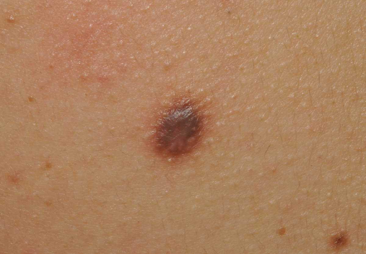
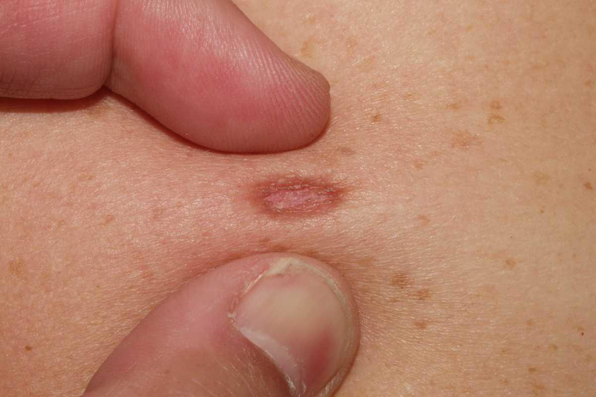
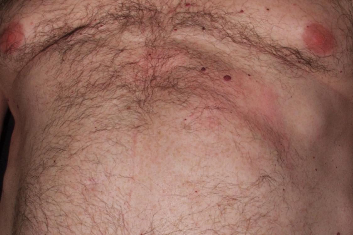
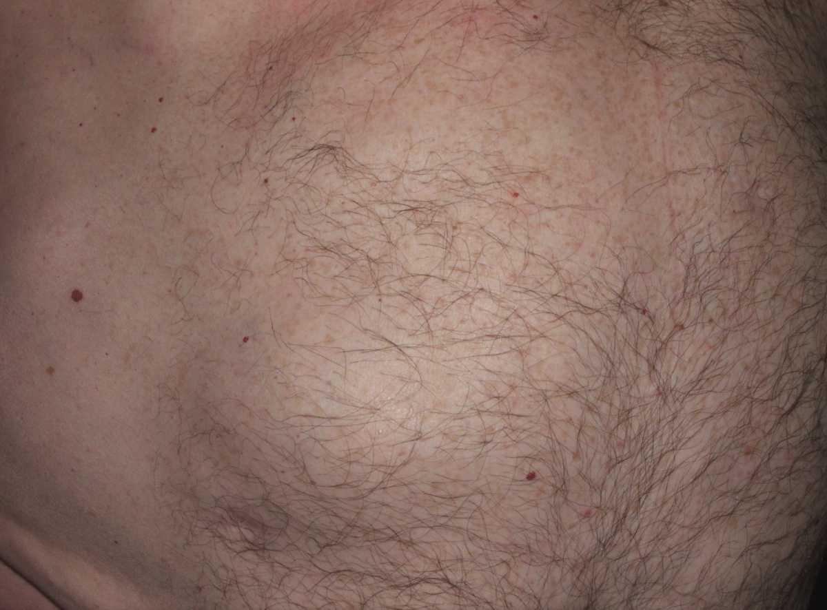
Imágenes tema

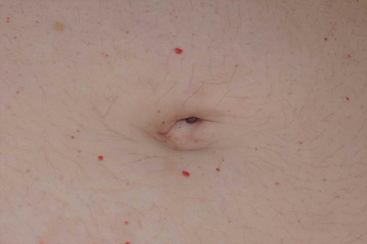
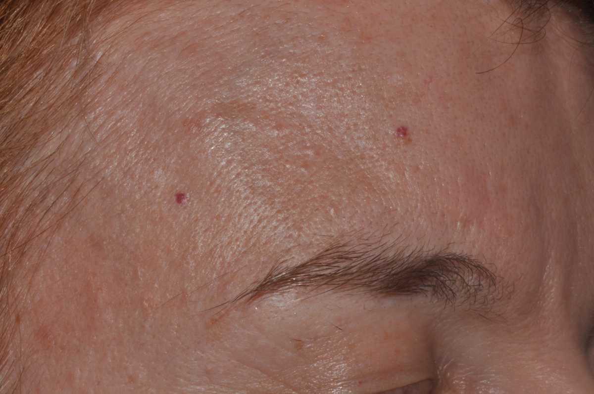
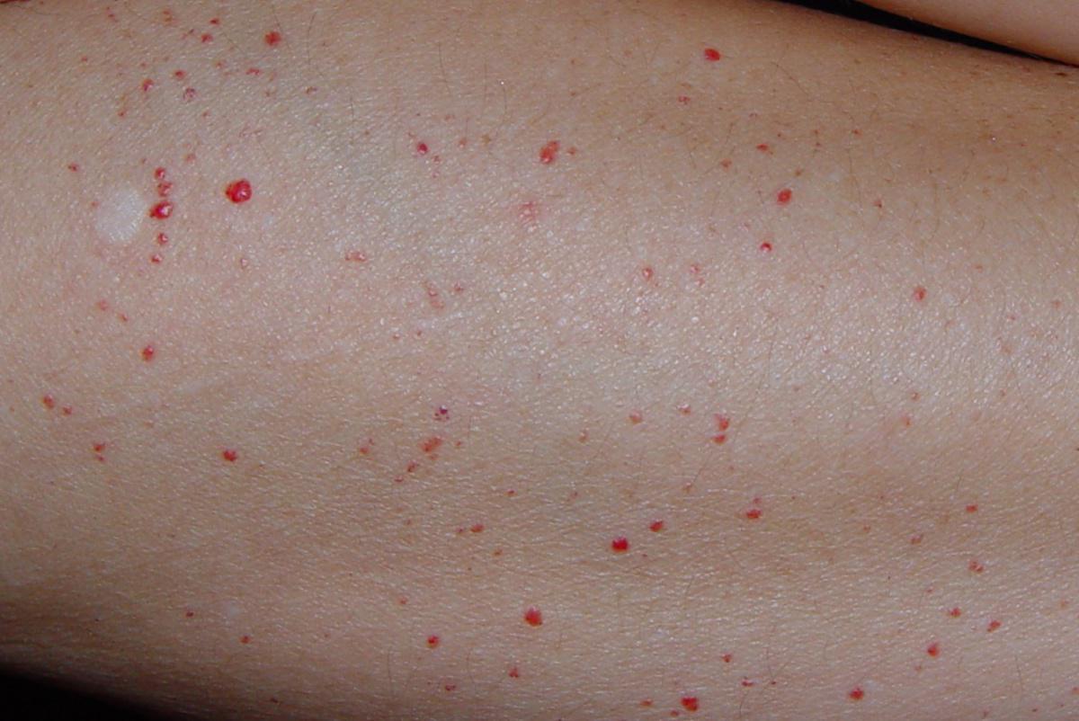
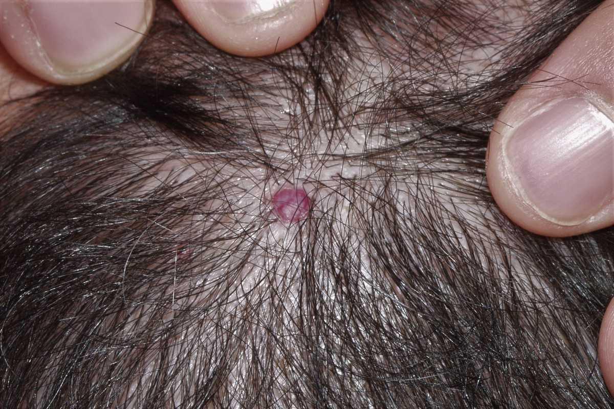
8. Bibliografía
-Pariser RJ. Benign neoplasms of the skin. Med Clin North Am. 1998; 82:1285-307.
-Bastiaens M, Hoefnagel J, Westendorp R, Vermeer BJ, Bouwes Bavinck JN. Solar lentigines are strongly related to sun exposure in contrast to ephelides. Pigment Cell Res. 2004; 17:225-9.
-Situm M, Bulat V, Buljan M, Puljiz Z, Situm V, Bolanca Z. Senile lentigo -cosmetic or medical issue of the elderly population. Coll Antropol. 2010; 34 Suppl 2:85-8.
-Ozbagcivan O, Akarsu S, Ikiz N, Semiz F, Fetil E. Dermoscopic Differentiation of Facial Lentigo Maligna from Pigmented Actinic Keratosis and Solar Lentigines. Acta Dermatovenerol Croat. 2019; 27:146-152.
-Krueger L, Saizan A, Stein JA, Elbuluk N. Dermoscopy of acquired pigmentary disorders: a comprehensive review. Int J Dermatol. 2022; 61(1):7-19.
-Tanaka M, Sawada M, Kobayashi K. Key points in dermoscopic differentiation between lentigo maligna and solar lentigo. J Dermatol. 2011; 38:53-8.
-Bagatin E, Gonçalves HS, Sato M, Almeida LMC, Miot HA. Comparable efficacy of adapalene 0.3% gel and tretinoin 0.05% cream as treatment for cutaneous photoaging. Eur J Dermatol. 2018; 28:343-350.
-Mukovozov I, Roesler J, Kashetsky N, Gregory A. Treatment of Lentigines: A Systematic Review. Dermatol Surg. 2023; 49(1):17-24.
Jackson JM, Alexis A, Berman B, Berson DS, Taylor S, Weiss JS. Current Understanding of Seborrheic Keratosis: Prevalence, Etiology, Clinical Presentation, Diagnosis, and Management. J Drugs Dermatol. 2015; 14:1119-25.
-Braun RP, Ludwig S, Marghoob AA. Differential Diagnosis of Seborrheic Keratosis: Clinical and Dermoscopic Features. J Drugs Dermatol. 2017; 16:835-84.
-Barthelmann S, Butsch F, Lang BM, Stege H, Großmann B, Schepler H, Grabbe S. Seborrheic keratosis. J Dtsch Dermatol Ges. 2023; 21(3):265-277.
-Wollina U. Recent advances in managing and understanding seborrheic keratosis. F1000Res. 2019;8. pii: F1000 Faculty Rev-1520.
-Hafner C, Hafner H, Groesser L. Genetic basis of seborrheic keratosis and pidermal nevi. Pathologe. 2014; 35:413-23.
-Salerni G, Alonso C, Gorosito M, Fernández-Bussy R. Seborrheic keratosis-like melanoma. J Am Acad Dermatol. 2015; 72(1 Suppl):S53-5.
-Karadag AS, Parish LC. The status of the seborrheic keratosis. Clin Dermatol. 2018; 36:275-277.
-Minagawa A. Dermoscopy-pathology relationship in seborrheic keratosis. J Dermatol. 2017; 44:518-524.
-Papageorgiou C, Spyridis I, Manoli SM, Busila I, Nasturica IE, Lallas K, Panagopoulou A, Papadimitriou I, Sideris N, Gentsidi T, Gonzalez-Cuevas R, Ilieva A, Ioannides D, Apalla Z, Lallas A. Accuracy of dermoscopic criteria for the differential diagnosis between irritated seborrheic keratosis and squamous cell carcinoma. J Am Acad Dermatol. 2020. pii: S0190-9622(20)30227-9.
-Zaresharifi S, Robati RM, Dadkhahfar S, Forouzanfar MM, Zaresharifi N. Efficacy and safety of cryotherapy, electrodesiccation, CO2 laser, and Er:YAG laser in the treatment of seborrheic keratosis. Dermatol Ther. 2021; 34(5):e15083.
-Jackson JM, Alexis A, Berman B, Berson DS, Taylor S, Weiss JS. Current Understanding of Seborrheic Keratosis: Prevalence, Etiology, Clinical Presentation, Diagnosis, and Management. J Drugs Dermatol. 2015; 14:1119-25.
-Kao S, Kiss A, Efimova T, Friedman AJ. Managing Seborrheic Keratosis: Evolving Strategies and Optimal Therapeutic Outcomes. J Drugs Dermatol. 2018; 17:933-940.
-Agrawal D, Adil M, Amin SS, Mohtashim M, Bansal R, Tabassum H. Comparison of efficacy & safety of 30% hydrogen peroxide with 50% trichloroacetic acid in seborrheic keratosis: a randomized controlled study. G Ital Dermatol Venereol. 2020. doi: 10.23736/S0392-0488.20.06518-9.
-Kwiek B, Schwartz RA. Keratoacanthoma (KA): An update and review. J Am Acad Dermatol. 2016; 74:1220-33.
–Savage JA, Maize JC Sr. Keratoacanthoma clinical behavior: a systematic review. Am J Dermatopathol. 2014; 36:422-9.
-Gardner HL, Cavanaugh RP. What is your diagnosis? Keratoacanthoma. J Am Vet Med Assoc. 2014; 244:1031-2.
-Zito PM, Scharf R. En: Keratoacanthoma. StatPearls [Internet]. Treasure Island (FL): StatPearls Publishing; 2018-.
-Takai T. Advances in histopathological diagnosis of keratoacanthoma. J Dermatol. 2017; 44:304-314.
-Goette DK, Odom RB, Arrott JW, Diakon NC, Horn RT Jr. Treatment of keratoacanthoma with topical application of fluorouracil. Arch Dermatol. 1982; 118:309-11.
-Kiss N, Avci P, Bánvölgyi A, Lőrincz K, Szakonyi J, Gyöngyösi N, Fésűs L, Lee G, Wikonkál N. Intralesional therapy for the treatment of keratoacanthoma. Dermatol Ther. 2019; 32(3):e12872.
-Moss M, Weber E, Hoverson K, Montemarano AD. Management of Keratoacanthoma: 157 Tumors Treated With Surgery or Intralesional Methotrexate. Dermatol Surg. 2019; 45:877-883.
-Melton JL, Nelson BR, Stough DB, Brown MD, Swanson NA, Johnson TM. Treatment of keratoacanthomas with intralesional methotrexate. J Am Acad Dermatol. 1991; 25(6 Pt 1):1017-23.
-Annest NM, VanBeek MJ, Arpey CJ, Whitaker DC. Intralesional methotrexate treatment for keratoacanthoma tumors: a retrospective study and review of the literature. J Am Acad Dermatol. 2007; 56:989-93.
-Gaitanis G, Bassukas ID. Cryosurgery, Intralesional Methotrexate and Imiquimod for Keratoacanthoma: Tuning the Combination. Case Rep Dermatol Med. 2019;2019:3489748.
-Sami N, Bussian A. Acitretin induces remission in generalized eruptive keratoacanthoma of Grzybowski. Int J Dermatol. 2015; 54:e67-9
-Vandeweyer E. Cutaneous cysts: a plea for systematic analysis. Acta Chir Belg. 2003; 103:507-10
-Terada T. Squamous cell carcinoma originated from an epidermal cyst. Int J Clin Exp Pathol. 2012; 5:479-81.
-Whittle C, Silva-Hirschberg C, Loyola K, Fajre X, Andrews A. Ultrasonographic Spectrum of Cutaneous Cysts With Stratified Squamous Epithelium in Pediatric Dermatology: Pictorial Essay. J Ultrasound Med. 2023; 42(4):923-930.
-Jenkins JR. Dermal cysts: a dermatopathological perspective and histological reappraisal. J Cutan Pathol. 2007; 34:815-29.
-Nakamura T. Comparative immunohistochemical analyses on the modes of cell death/keratinization in epidermal cyst, trichilemmal cyst, and pilomatricoma. Am J Dermatopathol. 2011; 33:78-83.
-Anand N, Kaur R, Saxena S, Bhardwaj N. Benign fibrous histiocytoma of the lower lip. J Oral Maxillofac Pathol. 2020; 24(Suppl 1):S97-S100.
-Queirós C, Uva L, Soares de Almeida L, Filipe P. Multiple eruptive dermatofibromas associated with pregnancy- a case and literature review. Dermatol Online J. 2019; 25(5). pii: 13030/qt29d3q6p1.
-Alves JV, Matos DM, Barreiros HF, Bártolo EA. Variants of dermatofibroma -a histopathological study. An Bras Dermatol. 2014; 89:472-7.
–Mohanty A, Mishra P, Kumar H, Panda A. A rare presentation of benign fibrous histiocytoma in the maxilla. J Oral Maxillofac Pathol. 2020; 24(Suppl 1):S73-S76.
–Zaballos P. Dermoscopy of dermatofibromas: a prospective morphological study of 412 cases. Arch Dermatol. 2008; 144:75-83.
-Ferrari A, Argenziano G, Buccini P, Cota C, Sperduti I, De Simone P, Eibenschutz L, Silipo V, Zalaudek I, Catricalà C. Typical and atypical dermoscopic presentations of dermatofibroma. J Eur Acad Dermatol Venereol. 2013; 27:1375-80.
-Charifa A, Badri T. Lipomas, Pathology. In: StatPearls [Internet]. Treasure Island (FL): StatPearls Publishing; 2020-2020 Mar 20.
-Johnson CN, Ha AS, Chen E, Davidson D. Lipomatous Soft-tissue Tumors. J Am Acad Orthop Surg. 2018; 26:779-788.
-Knebel C, Neumann J, Schwaiger BJ, Karampinos DC, Pfeiffer D, Specht K, Lenze U, von Eisenhart-Rothe R, Rummeny EJ, Woertler K, Gersing AS. Differentiating atypical lipomatous tumors from lipomas with magnetic resonance imaging: a comparison with MDM2 gene amplification status. BMC Cancer. 2019; 19:309.
-Murphy R, Hague A, Srinivasan J. A review of forehead lipomas: Important tips for the training surgeon. Surgeon. 2019; 17:186-189.
-Choi S, Nam K, Roh MR, Chung KY, Oh BH. Advantages of A Hairline Incision for the Excision of Forehead Lipomas. Dermatol Surg. 2020. doi: 10.1097/DSS.0000000000002330.
-Morioka D, Sato N, Ohkubo F. Excision of Large Lipomas Using Tumescent Local Anesthesia. J Cutan Med Surg. 2016; 20:263-5.
-Léauté-Labrèze C, Harper JI, Hoeger PH. Infantile haemangioma. Lancet. 2017; 390(10089):85-94.
-Baselga Torres E, Bernabéu Wittel J, van Esso Arbolave DL, Febrer Bosch MI, Carrasco Sanz Á, de Lucas Laguna R, Del Pozo Losada J, Hernández Martín Á, Jiménez Montañés L, López Gutiérrez JC, Martín-Santiago A, Redondo Bellón P, Ruíz-Canela Cáceres J, Torrelo Fernández A, Vera Casaño Á, Vicente Villa MA. [Spanish consensus on infantile haemangioma]. An Pediatr (Barc). 2016; 85:256-265.
-Boull C, Maguiness SM. Congenital hemangiomas. Semin Cutan Med Surg. 2016; 35:124-7.
-Levin LE, Lauren CT. Multifocal vascular lesions. Semin Cutan Med Surg. 2016; 35:153-60.
-Stefanko NS, Cossio ML, Powell J, Blei F, Davies OMT, Frieden IJ, Garzon MC, Lauren CT, Maheshwari M, McCuaig CC, Metry D, Salman S, Drolet BA, Siegel DH. Natural history of PHACE syndrome: A survey of adults with PHACE. Pediatr Dermatol. 2019. doi: 10.1111/pde.13871.
-Frade F, Kadlub N, Soupre V, Cassier S, Audry G, Vazquez MP, Picard A. Du PELVIS au LUMBAR syndrome: à propos de 2 cas. Arch Pediatr. 2012; 19:55-8.
–Smith CJF, Friedlander SF, Guma M, Kavanaugh A, Chambers CD. Infantile Hemangiomas: An Updated Review on Risk Factors, Pathogenesis, and Treatment. Birth Defects Res. 2017; 109:809-815.
–Wu W, Wang H, Hao J, Gao Z, Li F, Chen Y. Therapeutic efficacy of propranolol for infantile hemangiomas. Oral Surg Oral Med Oral Pathol Oral Radiol. 2019. doi: 10.1016/j.oooo.2019.04.014.
-Putterman E, Wan J, Streicher JL, Yan AC. Evaluation of a modified outpatient model for using propranolol to treat infantile hemangiomas. Pediatr Dermatol. 2019. doi: 10.1111/pde.13843.
-Yu Z, Cai R, Chang L, Qiu Y, Chen X, Chen Q, Ma G, Jin Y, Lin X. Clinical and radiological outcomes of infantile hemangioma treated with oral propranolol: A long-term follow-up study. J Dermatol. 2019; 46:376-382.
-Muñoz-Garza FZ, Ríos M, Roé-Crespo E, Bernabeu-Wittel J, Montserrat-García MT, Puig L, Gich I, Baselga E. Efficacy and Safety of Topical Timolol for the Treatment of Infantile Hemangioma in the Early Proliferative Stage: A Randomized Clinical Trial. JAMA Dermatol. 2021; 157(5):583-587.
-Mannschreck DB, Huang AH, Lie E, Psoter K, Puttgen K. Topical timolol as adjunct therapy to shorten oral propranolol therapy for infantile hemangiomas. Pediatr Dermatol. 2019; 36:283-289.
-Ziad K, Badi J, Roaa Z, Emily AH. Laser treatment of infantile hemangioma. J Cosmet Dermatol. 2023; 22 Suppl 2:1-7.
-McCuaig CC. Update on classification and diagnosis of vascular malformations. Curr Opin Pediatr. 2017; 29:448-454.
-Sadick M, Müller-Wille R, Wildgruber M, Wohlgemuth WA. Vascular Anomalies (Part I): Classification and Diagnostics of Vascular Anomalies. Rofo. 2018; 190:825-835.
-Olivieri B, White CL, Restrepo R, McKeon B, Karakas SP, Lee EY. Low-Flow Vascular Malformation Pitfalls: From Clinical Examination to Practical Imaging Evaluation–Part 2, Venous Malformation Mimickers. AJR Am J Roentgenol. 2016; 206:952-62.
-Dasgupta R, Patel M. Venous malformations. Semin Pediatr Surg. 2014; 23:198-202.
–Lee BB. Venous malformation and haemangioma: differential diagnosis, diagnosis, natural history and consequences. Phlebology. 2013; 28 Suppl 1:176-87.
-Clemens RK, Baumann F, Husmann M, Meier TO, Thalhammer C, MacCallum G, Amann-Vesti BR, Alomari AI. Percutaneous sclerotherapy for spongiform venous malformations – analysis of patient-evaluated outcome and satisfaction. Vasa. 2017; 46:477-483.
-Hage AN, Chick JFB, Srinivasa RN, Bundy JJ, Chauhan NR, Acord M, Gemmete JJ. Treatment of Venous Malformations: The Data, Where We Are, and How It Is Done. Tech Vasc Interv Radiol. 2018; 21:45-54.
–Malic CC, Guilfoyle R, Courtemanche RJM, Arneja JS, Heran MKS, Courtemanche DJ. Lymphatic Malformation Architecture: Implications for Treatment With OK-432. J Craniofac Surg. 2017; 28:1721-1724.
-Kim T, Tafoya E, Chelliah MP, Lekwuttikarn R, Li J, Sarin KY, Teng J. Alterations of the MEK/ERK, BMP, and Wnt/β-catenin pathways detected in the blood of individuals with lymphatic malformations. PLoS One. 2019; 14:e0213872.
-Elluru RG, Balakrishnan K, Padua HM. Lymphatic malformations: diagnosis and management. Semin Pediatr Surg. 2014; 23:178-85.
-White CL, Olivieri B, Restrepo R, McKeon B, Karakas SP, Lee EY. Low-Flow Vascular Malformation Pitfalls: From Clinical Examination to Practical Imaging Evaluation–Part 1, Lymphatic Malformation Mimickers. AJR Am J Roentgenol. 2016; 206:940-51.
–Defnet AM, Bagrodia N, Hernandez SL, Gwilliam N, Kandel JJ. Pediatric lymphatic malformations: evolving understanding and therapeutic options. Pediatr Surg Int. 2016; 32:425-33.
-Strychowsky JE, Rahbar R, O’Hare MJ, Irace AL, Padua H, Trenor CC 3rd. Sirolimus as treatment for 19 patients with refractory cervicofacial lymphatic malformation. Laryngoscope. 2018; 128:269-276.
-Johnson AB, Richter GT. Vascular Anomalies. Clin Perinatol. 2018; 45:737-749.
-Cavazos R, Patil MS, Gowda SH, Iacobas I, Rosenberg T, Fernandes CJ, Pammi M. Sirolimus for vascular anomalies in the first year of life: a systematic review. J Perinatol. 2024; 44(8):1087-1097.
-Afsar FS, Ortac R. Acquired port-wine stain in a pediatric patient. J Cutan Med Surg. 2006; 10:151-3.
-Couto JA, Ayturk UM, Konczyk DJ, Goss JA, Huang AY, Hann S, Reeve JL, Liang MG, Bischoff J, Warman ML, Greene AK. A somatic GNA11 mutation is associated with extremity capillary malformation and overgrowth. Angiogenesis. 2017; 20:303-306.
-Lee JW, Chung HY. Capillary Malformations (Portwine Stains) of the Head and Neck: Natural History, Investigations, Laser, and Surgical Management. Otolaryngol Clin North Am. 2018; 51:197-211.
-Ch’ng S, Tan ST. Facial port-wine stains – clinical stratification and risks of neuro-ocular involvement. J Plast Reconstr Aesthet Surg. 2008; 61:889-93.
-Sivarajan V, Maclaren WM, Mackay IR. The effect of varying pulse duration, wavelength, spot size, and fluence on the response of previously treated capillary vascular malformations to pulsed-dye laser treatment. Ann Plast Surg. 2006; 57:25-32.
-Whitehead KJ, Smith MC, Li DY. Arteriovenous malformations and other vascular malformation syndromes. Cold Spring Harb Perspect Med. 2013; 3:a006635.
-Diaz O, Scranton R. Endovascular treatment of arteriovenous malformations. Handb Clin Neurol. 2016; 136:1311-7.
-Jin Y, Yang X, Hua C, Lin X, Chen H, Ma G, Wang T, Yu W. Ethanol Embolotherapy for the Management of Refractory Chronic Skin Ulcers Caused by Arteriovenous Malformations. J Vasc Interv Radiol. 2018; 29:107-113.
-Chelliah MP, Do HM, Zinn Z, Patel V, Jeng M, Khosla RK, Truong MT, Marqueling A, Teng JMC. Management of Complex Arteriovenous Malformations Using a Novel Combination Therapeutic Algorithm. JAMA Dermatol. 2018; 154:1316-1319.
-Khasnis A, Gokula RM. Spider nevus. J Postgrad Med. 2002; 48:307-9.
-Li H, Wang R, Méndez-Sánchez N, Peng Y, Guo X, Qi X. Impact of spider nevus and subcutaneous collateral vessel of chest/abdominal wall on outcomes of liver cirrhosis. Arch Med Sci. 2019; 15:434-448.
-Finn SM, Rowland M, Lawlor F, Kinsella W, Chan L, Byrne O, O’Mahony O, Bourke B. The significance of cutaneous spider naevi in children. Arch Dis Child. 2006; 91:604-5.
-Sharma S, Chandra S, Gupta S, Srivastava S. Heterogeneous conceptualization of etiopathogenesis: Oral pyogenic granuloma. Natl J Maxillofac Surg. 2019; 10:3-7.
-Jafarzadeh H, Sanatkhani M, Mohtasham N. Oral pyogenic granuloma: a review. J Oral Sci. 2006; 48:167-75.
–Canivell-Zabaleta M, Martin-Lozano G, Olmos-Juarez E, Fontillon-Alberdi M, Infante-Cossio P. Extragingival Pregnancy Pyogenic Granuloma on the Lip. J Craniofac Surg. 2018; 29:e49-e50.
-Mills SE, Cooper PH, Fechner RE. Lobular capillary hemangioma: the underlying lesion of pyogenic granuloma. A study of 73 cases from the oral and nasal mucous membranes. Am J Surg Pathol. 1980; 4:470-9.
–Benedetto C, Crasto D, Ettefagh L, Nami N. Development of Periungual Pyogenic Granuloma with Associated Paronychia Following Isotretinoin Therapy: A Case Report and a Review of the Literature. J Clin Aesthet Dermatol. 2019; 12:32-36.
-Lacouture M, Sibaud V. Toxic Side Effects of Targeted Therapies and Immunotherapies Affecting the Skin, Oral Mucosa, Hair, and Nails. Am J Clin Dermatol. 2018; 19(Suppl 1):31‐39.
-Paulsen IF, Venzo A, Bredgaard R. Pyogenic granuloma. Ugeskr Laeger. 2014; 176(10A). pii: V11130681.
-Patrice SJ, Wiss K, Mulliken JB. Pyogenic granuloma (lobular capillary hemangioma): a clinicopathologic study of 178 cases. Pediatr Dermatol. 1991; 8:267-76.
-Moshe M, Levi A, Ad-El D, Ben-Amitai D, Mimouni D, Didkovsky E, Feinmesser M, Lapidoth M. Malignant melanoma clinically mimicking pyogenic granuloma: comparison of clinical evaluation and histopathology. Melanoma Res. 2018; 28:363-367.
–Zeng H, Yang R, Ding Y. Use of a water-cooled Nd:YAG pulsed laser in the treatment of giant gingival pyogenic granulomas during pregnancy. J Stomatol Oral Maxillofac Surg. 2019 Jun 1. pii: S2468-7855(19)30155-7.
-Daruwalla SB, Ghate S, Dhurat R. Establishing the efficacy and safety of the novel use of common salt for the treatment of pyogenic granuloma. Clin Exp Dermatol. 2021; 46(7):1243-1247.
-Patra AC, Sil A, Ahmed SS, Rahaman S, Mondal N, Roy S, Datta A, Kaliyadan F, Panda S, Setia MS, Dogra S, Khandpur S, Hazra A, Das NK. Effectiveness and safety of 0.5% timolol solution in the treatment of pyogenic granuloma: A randomized, double-blind and placebo-controlled study. Indian J Dermatol Venereol Leprol. 2022; 88(4):500-508.
-James WD, Berger TB, Elston D, eds. Dermal and subcutaneous tumors: cherry angiomas. Andrew’s Diseases of the Skin: Clinical Dermatology. 11h ed. Philadelphia, Pa: WB Saunders; 2011. 574-619.