
Currículum
Tema 13. Vitíligo y otras dermatosis con cambios en el color de la piel
1. VITÍLIGO
0/3

2. OTRAS DERMATOSIS HIPOPIGMENTADAS
0/4
3. DERMATOSIS HIPERPIGMENTADAS
0/4
4. BIBLOGRAFIA
0/1
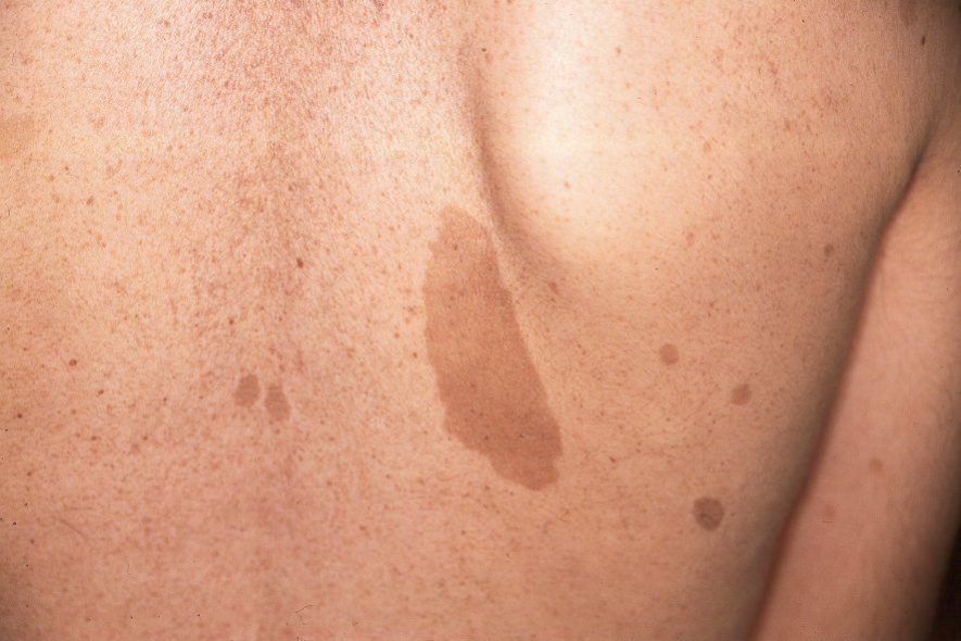
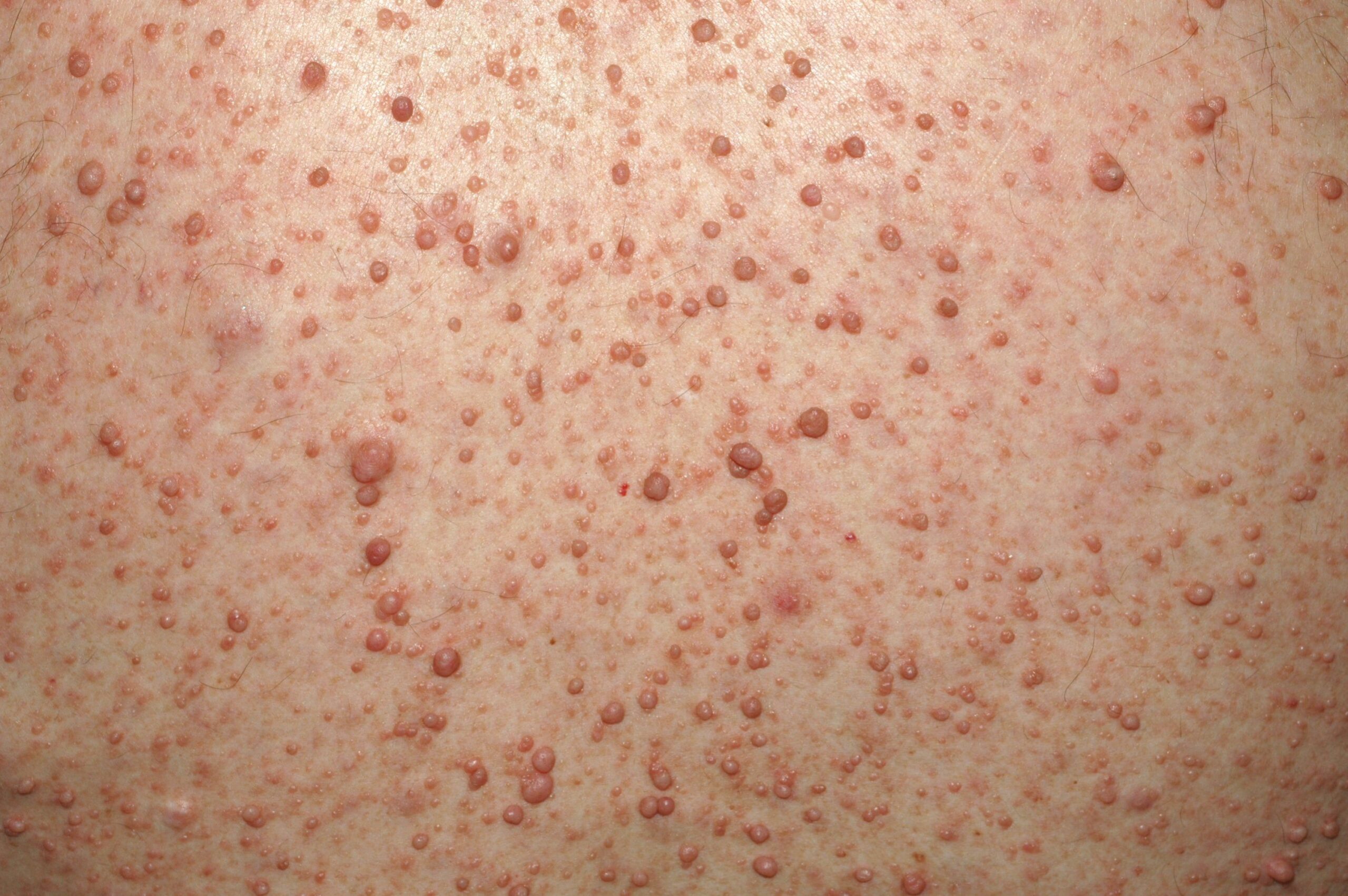
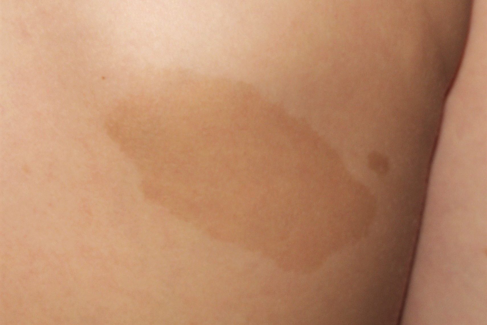
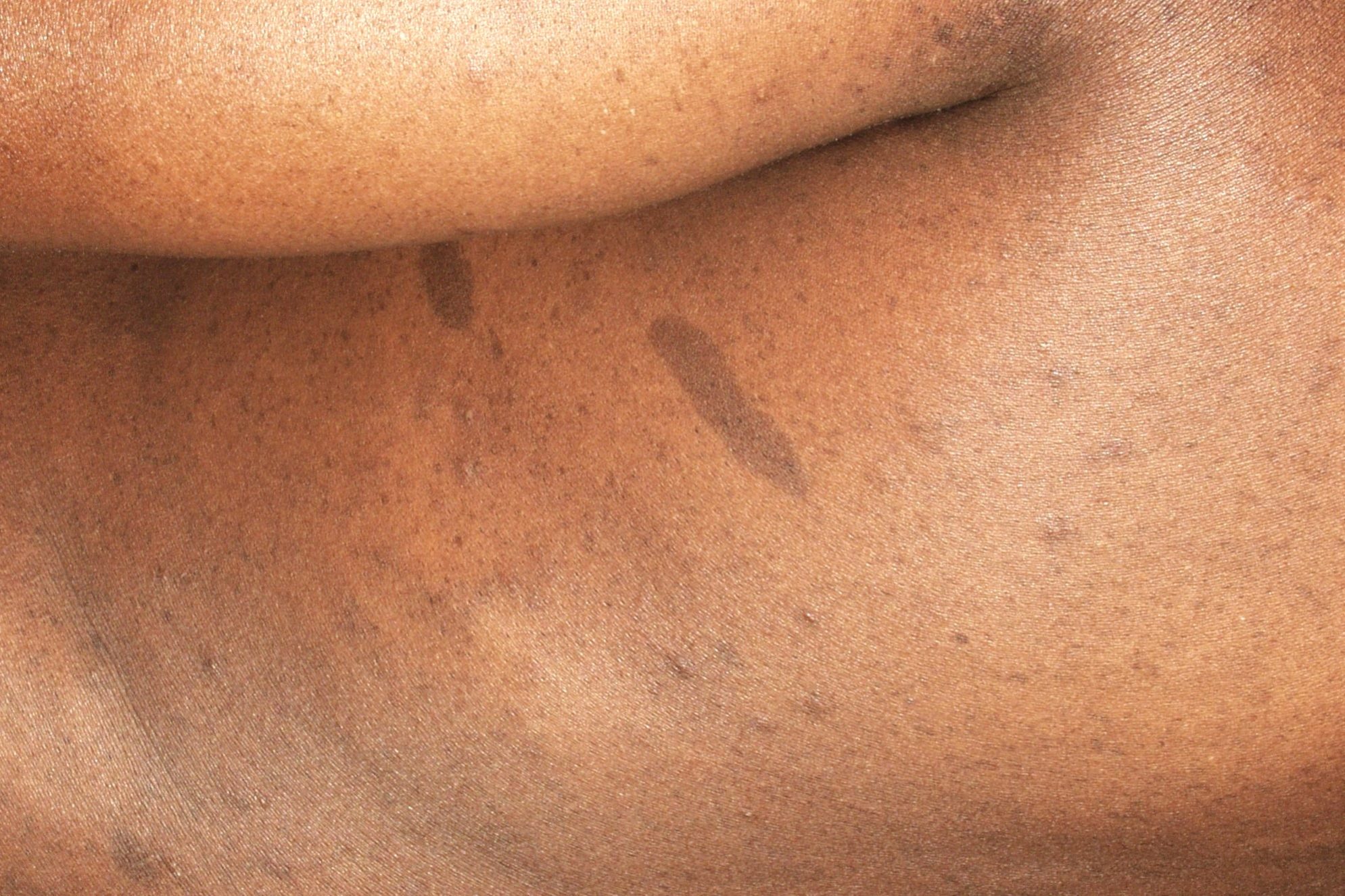
Imágenes tema

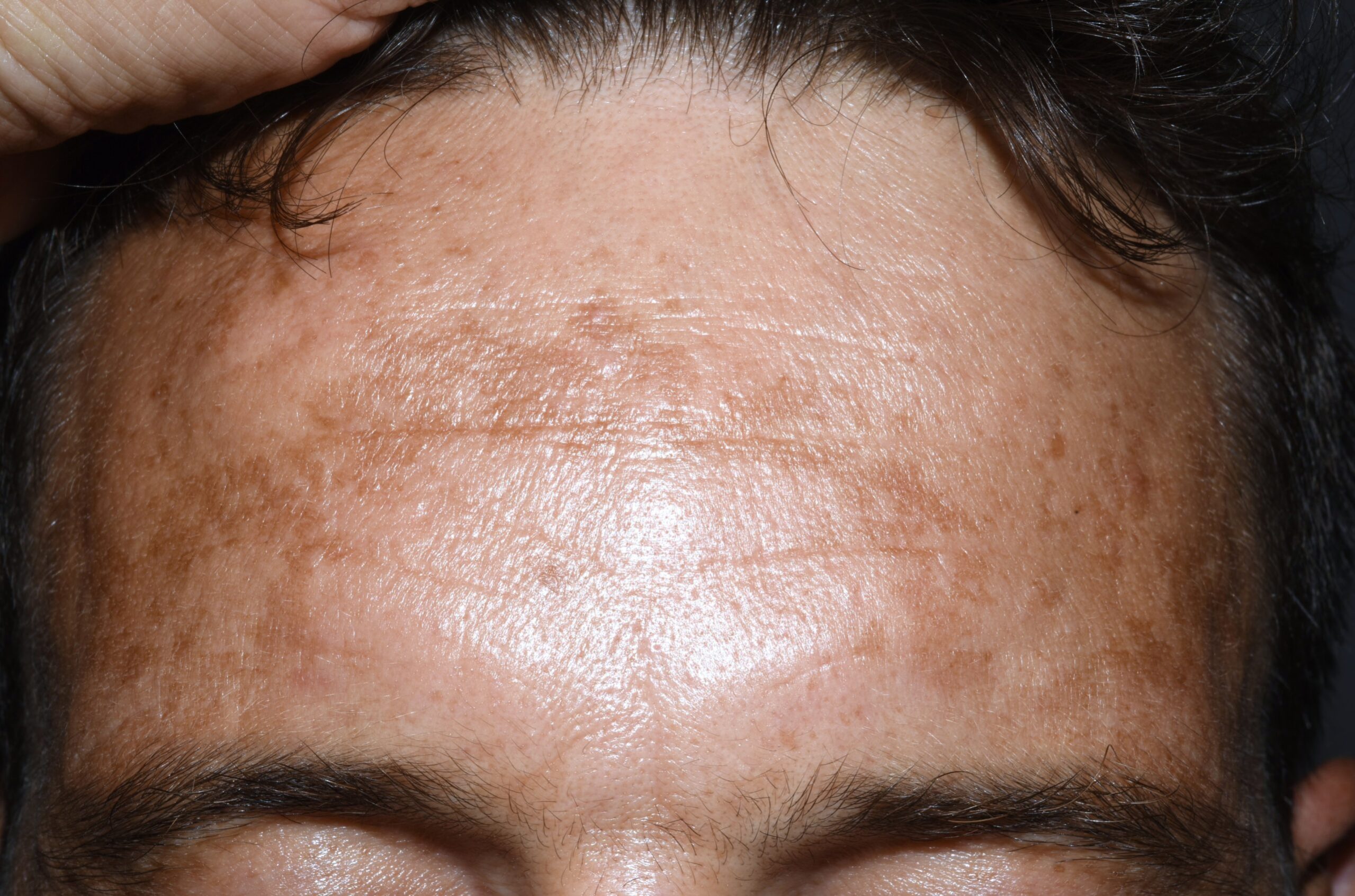
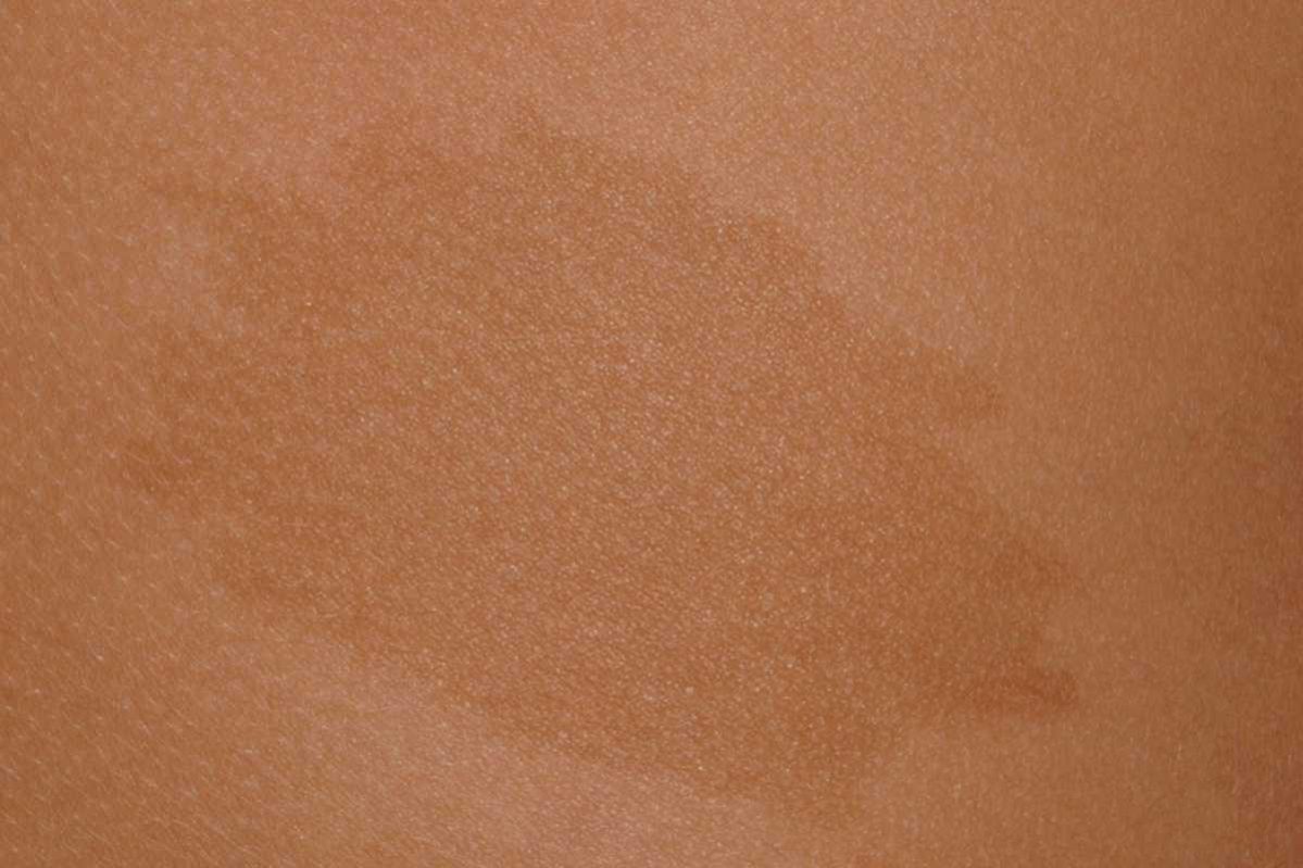
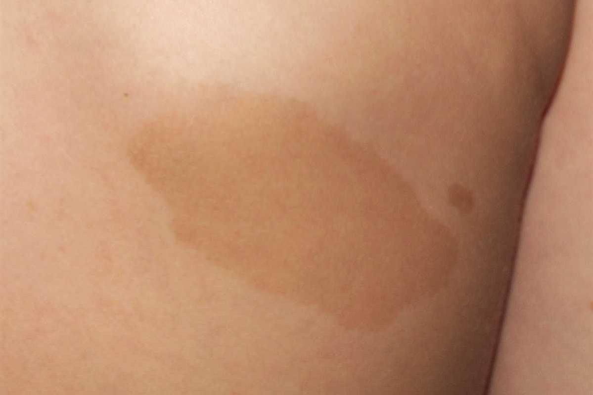
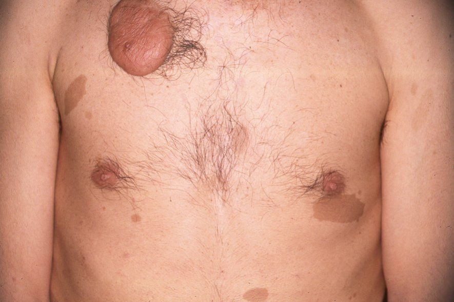
4.1. Bibliografía
4.1. Bibliografía
-Ahmed jan N, Masood S. Vitiligo. 2023 Aug 7. In: StatPearls [Internet]. Treasure Island (FL): StatPearls Publishing; 2025 Jan–.
-Albelowi LM, Alhazmi RM, Ibrahim S. The Pathogenesis and Management of Vitiligo. Cureus. 2024; 16(12):e75859.
-Haulrig MB, Idorn L, Skov L. Vitiligo. Ugeskr Laeger. 2025; 187(14):V10240662.
-Renert-Yuval Y, Desai S, Huang V, Walsh S, Rosmarin D, Silverberg N. A Review of the Vitiligo Literature to Standardize Expression of Disease Severity. J Drugs Dermatol. 2024; 23(10):842-846.
-Kussainova A, Kassym L, Akhmetova A, Glushkova N, Sabirov U, Adilgozhina S, Tuleutayeva R, Semenova Y. Vitiligo and anxiety: A systematic review and meta-analysis. PLoS One. 2020; 15(11):e0241445.
-Ezzedine K, Eleftheriadou V, Jones H, Bibeau K, Kuo FI, Sturm D, Pandya AG. Psychosocial Effects of Vitiligo: A Systematic Literature Review. Am J Clin Dermatol. 2021; 22(6):757-774.
-Tarlé RG, Nascimento LM, Mira MT, Castro CC. Vitiligo–part 1. An Bras Dermatol. 2014; 89:461-70.
-Spritz RA, Santorico SA. The Genetic Basis of Vitiligo. J Invest Dermatol. 2021; 141(2):265-273.
-Roberts GHL, Paul S, Yorgov D, Santorico SA, Spritz RA. Family Clustering of Autoimmune Vitiligo Results Principally from Polygenic Inheritance of Common Risk Alleles. Am J Hum Genet. 2019; 105(2):364-372.
-Alkhateeb A, Fain PR, Thody A, Bennett DC, Spritz RA. Epidemiology of vitiligo and associated autoimmune diseases in Caucasian probands and their families. Pigment Cell Res. 2003; 16(3):208-14.
-Laberge G, Mailloux CM, Gowan K, Holland P, Bennett DC, Fain PR, Spritz RA. Early disease onset and increased risk of other autoimmune diseases in familial generalized vitiligo. Pigment Cell Res. 2005; 18(4):300-5.
-Speeckaert R, van Geel N. Vitiligo: An Update on Pathophysiology and Treatment Options. Am J Clin Dermatol. 2017; 18(6):733-744.
-Leung AKC, Lam JM, Leong KF, Hon KL. Vitiligo: An Updated Narrative Review. Curr Pediatr Rev. 2021; 17(2):76-91.
-Roberts GHL, Santorico SA, Spritz RA. The genetic architecture of vitiligo. Pigment Cell Melanoma Res. 2020; 33(1):8-15.
-Spritz RA, Santorico SA. The Genetic Basis of Vitiligo. J Invest Dermatol. 2021; 141(2):265-273.
-Okamura K, Suzuki T. Genetics and epigenetics in vitiligo. J Dermatol Sci. 2025; 117(3):45-51.
-Yamaguchi HL, Yamaguchi Y, Peeva E. Pathogenesis of Alopecia Areata and Vitiligo: Commonalities and Differences. Int J Mol Sci. 2024; 25(8):4409.
-Frisoli ML, Essien K, Harris JE. Vitiligo: Mechanisms of Pathogenesis and Treatment. Annu Rev Immunol. 2020; 38:621-648.
-Qi F, Liu F, Gao L. Janus Kinase Inhibitors in the Treatment of Vitiligo: A Review. Front Immunol. 2021; 12:790125.
-Pathak GN, Tan IJ, Bai G, Dhillon J, Rao BK. Vitiligo: From mechanisms of disease to treatable pathways. Skin Health Dis. 2024; 4(6):e460.
-Prajapati VH, Lui H, Miller-Monthrope Y, Ringuet J, Turchin I, Hong HC, Lynde C, Papp KA, Yeung J, Gooderham MJ. Canadian Consensus Guidelines for the Management of Vitiligo. Dermatol Ther (Heidelb). 2025; 15(6):1351-1369.
-Böhm M, Tanew A. Vitiligo. J Dtsch Dermatol Ges. 2025; 23(8):968-987.
-Searle T, Al-Niaimi F, Ali FR. Vitiligo: an update on systemic treatments. Clin Exp Dermatol. 2021; 46(2):248-258.
-Seneschal J, Speeckaert R, Taïeb A, Wolkerstorfer A, Passeron T, Pandya AG, Lim HW, Ezzedine K, Zhou Y, Xiang F, Thng S, Tanemura A, Suzuki T, Rosmarin D, Rodrigues M, Raboobee N, Pliszewski G, Parsad D, Oiso N, Monteiro P, Meurant JM, Maquignon N, Lui H, Le Poole C, Leone G, Lee AY, Lan E, Katayama I, Huggins R, Oh SH, Harris JE, Hamzavi IH, Gupta S, Grimes P, Goh BK, Ghia D, Esmat S, Eleftheriadou V, Böhm M, Benzekri L, Bekkenk M, Bae JM, Alomar A, Abdallah M, Picardo M, van Geel N. Worldwide expert recommendations for the diagnosis and management of vitiligo: Position statement from the international Vitiligo Task Force-Part 2: Specific treatment recommendations. J Eur Acad Dermatol Venereol. 2023; 37(11):2185-2195.
-Das K, Sarka.r R, Sinha S. Recent Advances in Management of Vitiligo. Indian J Dermatol. 2025; 70(4):201-207.
-Renert-Yuval Y, Ezzedine K, Grimes P, Rosmarin D, Eichenfield LF, Castelo-Soccio L, Huang V, Desai SR, Walsh S, Silverberg JI, Paller AS, Rodrigues M, Weingarten M, Narla S, Gardner J, Siegel M, Ibad S, Silverberg NB. Expert Recommendations on Use of Topical Therapeutics for Vitiligo in Pediatric, Adolescent, and Young Adult Patients. JAMA Dermatol. 2024; 160(4):453-461.
-Leung AKC, Lam JM, Leong KF, Hon KL. Vitiligo: An Updated Narrative Review. Curr Pediatr Rev. 2021; 17(2):76-91.
-Hu W, Lin F, Lei J, Xu AE. Impacts of exposure to topical calcineurin inhibitors on metabolism in vitiligo infants. Pediatr Res. 2023; 93(3):661-665.
-Dang YP, Li Q, Shi F, Yuan XY, Liu W. Effect of topical calcineurin inhibitors as monotherapy or combined with phototherapy for vitiligo treatment: a meta-analysis. Dermatol Ther. 2016; 29(2):126-33.
-Radakovic S, Breier-Maly J, Konschitzky R, Kittler H, Sator P, Hoenigsmann H, Tanew A. Response of vitiligo to once- vs. twice-daily topical tacrolimus: a controlled prospective, randomized, observer-blinded trial. J Eur Acad Dermatol Venereol. 2009; 23(8):951-3.
-Duplaine A, Tannous J, Seneschal J, Ezzedine K, Passeron T, Dupin N, Quereux G, Beylot-Barry M, Chosidow O, Guillot B; Centre of Evidence of Société Française de Dermatologie. Value of tacrolimus 0.1% in the treatment of vitiligo in the era of targeted therapy. Ann Dermatol Venereol. 2025; 152(2):103352.
-Roy P, Saha SK, Paul PC, Reza AK, Nandi AK, Sultana S, Saha S, Akhter SM, Khatun S, Habibunnahar M. Effectiveness of Topical Corticosteroid, Topical Calcineurin Inhibitors and Combination of Them in the Treatment of Vitiligo. Mymensingh Med J. 2016 Oct;25(4):620-627.
-Alqifari SF, Gari MH, Guo JJ, Alamin S, Esmail AK, Esmail AK, Hamad HR, Aljabri A, Alatawi AM, Albishi LA, Alraddadi MO, Hetta HF. Descriptive Analysis of Reported Adverse Events Associated with Vitiligo Medications Using FDA Adverse Event Reporting System (FAERS) Databases 2013-2023. Diseases. 2025; 13(7):208.
-Inoue S, Suzuki T, Sano S, Katayama I. JAK inhibitors for the treatment of vitiligo. J Dermatol Sci. 2024; 113(3):86-92.
-Seidel P, Böhm M. Vitiligo – Update zu Pathogenese, Diagnostik und Therapie. Dermatologie (Heidelb). 2025; 76(3):168-178.
-Rosmarin D, Passeron T, Pandya AG, Grimes P, Harris JE, Desai SR, Lebwohl M, Ruer-Mulard M, Seneschal J, Wolkerstorfer A, Kornacki D, Sun K, Butler K, Ezzedine K; TRuE-V Study Group. Two Phase 3, Randomized, Controlled Trials of Ruxolitinib Cream for Vitiligo. N Engl J Med. 2022; 387(16):1445-1455).
-Ezzedine K, Peeva E, Yamaguchi Y, Cox LA, Banerjee A, Han G, Hamzavi I, Ganesan AK, Picardo M, Thaçi D, Harris JE, Bae JM, Tsukamoto K, Sinclair R, Pandya AG, Sloan A, Yu D, Gandhi K, Vincent MS, King B. Efficacy and safety of oral ritlecitinib for the treatment of active nonsegmental vitiligo: A randomized phase 2b clinical trial. J Am Acad Dermatol. 2023; 88(2):395-403.
-Gupta AK, Bamimore MA, Seneschal J, Piguet V, Talukder M. Relative Impact of Monotherapies for Vitiligo: A Network Meta-Analysis Study. J Cosmet Dermatol. 2025; 24(3):e70078.
-Ezzedine K, Eleftheriadou V, Whitton M, van Geel N. Vitiligo. Lancet. 2015; 386(9988):74-84.
-Hearn RMR, Kerr AC, Rahim KF, Ferguson J, Dawe RS. Incidence of skin cancers in 3867 patients treated with narrow-band ultraviolet B phototherapy. Br J Dermatol. 2008; 159(4):931-5.
-Bae JM, Ju HJ, Lee RW, Oh SH, Shin JH, Kang HY, Kang HY, Park JH, Kim HJ, Jeong KH, Lee HJ, Lee S, Kim DH, Lee DY, Kim YC, Choi GS, Kim KH, Park CJ, Choi CW; Korean Society of Vitiligo. Evaluation for Skin Cancer and Precancer in Patients With Vitiligo Treated With Long-term Narrowband UV-B Phototherapy. JAMA Dermatol. 2020; 156(5):529-537.
-Al Jasser M, Richer V, Ball N, Lui H, Zhou Y. Photolichenoid papules within vitiligo induced by narrowband UVB phototherapy. J Eur Acad Dermatol Venereol. 2016; 30(8):1428-9.
-Luo L, Huang J, Fu C, Hu Y, Chen J, Jiang L, Zeng Q. The efficacy of combined phototherapy with topical therapy in vitiligo: a network meta-analysis. J Dermatolog Treat. 2025; 36(1):2483808.
-Dang YP, Li Q, Shi F, Yuan XY, Liu W. Effect of topical calcineurin inhibitors as monotherapy or combined with phototherapy for vitiligo treatment: a meta-analysis. Dermatol Ther. 2016; 29(2):126-33.
-Suo DF, Zeng SW, Meng LH. 308 nm excimer laser and tacrolimus ointment in the treatment of facial vitiligo: a systematic review and meta-analysis. Lasers Med Sci. 2024; 39(1):90).
-Ninama K, Mahajan R, Baxi D, Patil RS, Marfatia Y. A comparative study of the efficacy of potent topical corticosteroids per se versus combination of potent topical corticosteroids with fractional CO2 laser in the management of vitiligo vulgaris. J Cutan Aesthet Surg. 2025; 18(1):42-47.
-Qian YT, Liu JW, Ma DL. A 5-week low-dose prednisolone regimen for progressive vitiligo: efficacy and safety in a single-center cohort study. Ann Med. 2025; 57(1):2543523.
-Feng Y, Lu Y. Advances in vitiligo: Update on therapeutic targets. Front Immunol. 2022; 13:986918.
-van Geel N, Lommerts J, Bekkenk M, Wolkerstorfer A, Prinsen CAC, Eleftheriadou V, Taïeb A, Picardo M, Ezzedine K, Speeckaert R. Development and validation of the vitiligo extent score (VES): an international collaborative initiative. J Invest Dermatol. 2016; 136(5):978-84.
-Bidaki R, Majidi N, Moghadam Ahmadi A, Bakhshi H, Sadr Mohammadi R, Mostafavi SA, Kazemi Arababadi M, Hadavi M, Mirzaei A. Vitiligo and social acceptance. Clin Cosmet Investig Dermatol. 2018; 11:383-6.
-Mosher DB, Parrish JA, Fitzpatrick TB. Monobenzylether of hydroquinone. A retrospective study of treatment of 18 vitiligo patients and a review of the literature. Br J Dermatol. 1977; 97(6):669-79.
-van Geel N, Depaepe L, Speeckaert R. Laser (755 nm) and cryotherapy as depigmentation treatments for vitiligo: a comparative study. J Eur Acad Dermatol Venereol. 2015; 29(6):1121-7.
-Majid I, Imran S. Depigmentation therapy with Q-switched Nd: YAG laser in universal vitiligo. J Cutan Aesthet Surg. 2013; 6(2):93-6.
-Leung AK, Barankin B, Lam JM, Leong KF, Hon KL. Tinea versicolor: an updated review. Drugs Context. 2022; 11:2022-9.
-Park M, Park S, Jung WH. Skin Commensal Fungus Malassezia and Its Lipases. J Microbiol Biotechnol. 2021; 31(5):637-644.
-Saunte DML, Gaitanis G, Hay RJ. Malassezia-Associated Skin Diseases, the Use of Diagnostics and Treatment. Front Cell Infect Microbiol. 2020; 10:112.
-Karray M, McKinney WP. Tinea Versicolor. In: StatPearls [Internet]. Treasure Island (FL): StatPearls Publishing; 2018 Jan-.
-Karray M, McKinney WP. Tinea Versicolor. 2024 Feb 12. In: StatPearls [Internet]. Treasure Island (FL): StatPearls Publishing; 2025 Jan–.
-Kurniadi I, Hendra Wijaya W, Timotius KH. Malassezia virulence factors and their role in dermatological disorders. Acta Dermatovenerol Alp Pannonica Adriat. 2022; 31(2):65-70.
-Chueachavalit C, Meephansan J, Payungporn S, Sawaswong V, Chanchaem P, Wongpiyabovorn J, Thio HB. Comparison of Malassezia spp. colonization between human skin exposed to high- and low-ambient air pollution. Exp Dermatol. 2022; 31(9):1454-1461.
-Veraldi S, Aromolo IF, Germiniasi FS, Nazzaro G. Erythrasmoid pityriasis versicolor: three case reports and review of the literature. Dermatol Reports. 2024; 17(1):9976.
-Andersen PL, Andersen SML, Henning MAS, Svendsen MB, Astvad K, Hald M, Blomberg M, Saunte DML. Pityriasis versicolor. Ugeskr Laeger. 2025; 187(14):V10240670.
-Gupta AK, Wang T, Polla Ravi S, Mann A, Lincoln SA, Foreman HC, Bakotic WL. Epidemiology of Onychomycosis in the United States Characterized Using Molecular Methods, 2015-2024. J Fungi (Basel). 2024; 10(9):633.
-Marinello E, Piaserico S, Alaibac M. Atrophic pityriasis versicolor occurring in a patient with Sjögren’s syndrome. BMJ Case Rep. 2017; pii: bcr2016218108.
-Mathur M, Acharya P, Karki A, Kc N, Shah J. Dermoscopic pattern of pityriasis versicolor. Clin Cosmet Investig Dermatol. 2019; 12:303-309.
-Kaur I, Jakhar D, Singal A. Dermoscopy in the Evaluation of Pityriasis Versicolor: A Cross Sectional Study. Indian Dermatol Online J. 2019; 10(6):682-685.
-Vishwanath T, Kapadia FM, Mahajan SA, Kharkar VD, Chikhalkar SB, Gole PV, Iyer M, Shah AJ. Dermoscopic Perspective of Pityriasis Versicolor in a Cross-Sectional Study. Indian Dermatol Online J. 2024; 15(2):218-225.
-Vachiramon V, Thadanipon K. Postinflammatory hypopigmentation. Clin Exp Dermatol. 2011; 36:708-14.
-Madu PN, Syder N, Elbuluk N. Postinflammatory hypopigmentation: a comprehensive review of treatments. J Dermatolog Treat. 2022; 33(2):704-708.
-Kingston P, Jones M, Luu M. Common Causes of Hypopigmentation in Children. Pediatr Rev. 2025; 46(10):545-556.
–Rao M, Young K, Jackson-Cowan L, Kourosh A, Theodosakis N. Post-Inflammatory Hypopigmentation: Review of the Etiology, Clinical Manifestations, and Treatment Options. J Clin Med. 2023; 12(3):1243.
-Ramírez-Marín HA, Silverberg JI. Differences between pediatric and adult atopic dermatitis. Pediatr Dermatol. 2022; 39(3):345-353.
-Givler DN, Givler A. Pityriasis Alba. In: StatPearls [Internet]. Treasure Island (FL): StatPearls Publishing; 2018 Jan-.
-Givler DN, Saleh HM, Givler A. Pityriasis Alba. 2024 Jan 25. In: StatPearls [Internet]. Treasure Island (FL): StatPearls Publishing; 2025 Jan–.
-Jadotte YT, Janniger CK. Pityriasis alba revisited: perspectives on an enigmatic disorder of childhood. Cutis. 2011; 87:66-72.
-Miazek N, Michalek I, Pawlowska-Kisiel M, Olszewska M, Rudnicka L. Pityriasis Alba–Common Disease, Enigmatic Entity: Up-to-Date Review of the Literature. Pediatr Dermatol. 2015; 32:786-91.
-Lv Y, Gao Y, Lan N, Sun M, Zhang C, Gao J, Wang N, Yang X. Analysis of Epidemic Characteristics and Related Pathogenic Factors of 2726 Cases of Pityriasis Alba. Clin Cosmet Investig Dermatol. 2022; 15:203-209.
-Thomas IN, James JJ, Bala A, Mohan S, Dogiparthi S, Shanmugam NP Sr. Usage of Dermoscopy as an Effective Diagnostic Tool in Pityriasis Alba: A Prospective Observational Study Among Children in a Suburban Hospital in South India. Cureus. 2023; 15(6):e40271.
-Abdel-Wahab HM, Ragaie MH. Pityriasis alba: toward an effective treatment. J Dermatolog Treat. 2022; 33(4):2285-2289.
-Northrup H, Krueger DA. International Tuberous Sclerosis Complex Consensus Group. Tuberous sclerosis complex diagnostic criteria update: Recommendations of the 2012 Iinternational Tuberous Sclerosis Complex Consensus Conference. Pediatr. Neurol. 2013; 49:243-254.
-Lam HC, Nijmeh J, Henske EP. New developments in the genetics and pathogenesis of tumours in tuberous sclerosis complex. J Pathol. 2017; 241(2):219-225.
-Amorim BDB, Azulay D, Ramos-E-Silva M. Tuberous Sclerosis. Skinmed. 2021 Jun 1;19(3):179-185.
-Northrup H, Aronow ME, Bebin EM, Bissler J, Darling TN, de Vries PJ, Frost MD, Fuchs Z, Gosnell ES, Gupta N, Jansen AC, Jóźwiak S, Kingswood JC, Knilans TK, McCormack FX, Pounders A, Roberds SL, Rodriguez-Buritica DF, Roth J, Sampson JR, Sparagana S, Thiele EA, Weiner HL, Wheless JW, Towbin AJ, Krueger DA; International Tuberous Sclerosis Complex Consensus Group. Updated International Tuberous Sclerosis Complex Diagnostic Criteria and Surveillance and Management Recommendations. Pediatr Neurol. 2021; 123:50-66.
–Takahashi A, Hattori S, Sakai E, Yang L, Katayama I, Fujimoto M, Wataya-Kaneda M. Distribution of hypomelanotic macules in tuberous sclerosis complex: A retrospective cohort study. J Am Acad Dermatol. 2022; 87(1):237-240.
-Jóźwiak S, Schwartz RA, Janniger CK, Michałowicz R, Chmielik J. Skin lesions in children with tuberous sclerosis complex: their prevalence, natural course, and diagnostic significance. Int J Dermatol. 1998; 37(12):911-7.
-Dutta A, Ghosh SK, Mandal RK. Facial Angiofibromas. Indian Pediatr. 2015;52(7):634.
-Tyburczy ME, Wang JA, Li S, Thangapazham R, Chekaluk Y, Moss J, Kwiatkowski DJ, Darling TN. Sun exposure causes somatic second-hit mutations and angiofibroma development in tuberous sclerosis complex. Hum Mol Genet. 2014; 23(8):2023-9.
-Liebman JJ, Nigro LC, Matthews MS. Koenen tumors in tuberous sclerosis: a review and clinical considerations for treatment. Ann Plast Surg. 2014; 73(6):721-2.
-Takahashi A, Hattori S, Sakai E, Yang L, Katayama I, Fujimoto M, Wataya-Kaneda M. Distribution of shagreen patches and fibrous cephalic plaques in patients with tuberous sclerosis complex: A retrospective cohort study. J Am Acad Dermatol. 2024; 90(4):849-852.
-Bongiorno MA, Nathan N, Oyerinde O, Wang JA, Lee CR, Brown GT, Moss J, Darling TN. Clinical Characteristics of Connective Tissue Nevi in Tuberous Sclerosis Complex With Special Emphasis on Shagreen Patches. JAMA Dermatol. 2017; 153(7):660-665.
-Neema S, Mukherjee S, Battula S. Cephalic Fibrous Plaque in Tuberous Sclerosis. Indian Pediatr. 2023; 60(4):327.
–Torrelo A, Hadj-Rabia S, Colmenero I, Piston R, Sybert VP, Hilari-Carbonell H, Hernández-Martín A, Ferreres JC, Vañó-Galván S, Azorín D, de Salamanca JE, Requena L, Bodemer C, Happle R, García-Patos V, Fraitag S. Folliculocystic and collagen hamartoma of tuberous sclerosis complex. J Am Acad Dermatol. 2012; 66(4):617-21.
-Islam MP. Tuberous Sclerosis Complex. Semin Pediatr Neurol. 2021; 37:100875.
-Aronica E, Specchio N, Luinenburg MJ, Curatolo P. Epileptogenesis in tuberous sclerosis complex-related developmental and epileptic encephalopathy. Brain. 2023; 146(7):2694-2710.
-Kotulska K, Kwiatkowski DJ, Curatolo P, Weschke B, Riney K, Jansen F, Feucht M, Krsek P, Nabbout R, Jansen AC, Wojdan K, Sijko K, Głowacka-Walas J, Borkowska J, Sadowski K, Domańska-Pakieła D, Moavero R, Hertzberg C, Hulshof H, Scholl T, Benova B, Aronica E, de Ridder J, Lagae L, Jóźwiak S; EPISTOP Investigators. Prevention of Epilepsy in Infants with Tuberous Sclerosis Complex in the EPISTOP Trial. Ann Neurol. 2021; 89(2):304-314.
–Marlais M, Mekahli D. Tuberous sclerosis complex-associated kidney disease in children. Pediatr Nephrol. 2025; 40(6):1871-1877.
-Gupta S, Kang HC, Faria SC, Choyke PL, Kundra V. Tuberous Sclerosis Complex (TSC): Renal and Extrarenal Imaging. Acad Radiol. 2022; 29(3):439-449.
-Prelevic V, Juric I, Bevc S, Marcun-Varda N, Aleckovic-Halilovic M, Mesic E, Bilic H, Grujicic M, Zabic I, Josipovic J, Vujicic B, Marinaki S, Simic-Ogrizovic S, Milinkovic M, Azasevac T, Idrizi A, Arnol M, Radunovic D, Antunovic T, Jukic NB. Association of multiple retinal nodular hamartomas and “confetti” skin lesions with end-stage renal disease in patients with tuberous sclerosis. Int Urol Nephrol. 2023; 55(2):477-482.
-Dagge A, Silva LA, Jorge S, Nogueira E, Rebelo M, Pinto L. Fetal Tuberous Sclerosis: Sirolimus for the Treatment of Fetal rhabdomyoma. Fetal Pediatr Pathol. 2022; 41(5):800-806.
-Racioppi G, Proietti Checchi M, Sforza G, Voci A, Mazzone L, Valeriani M, Moavero R. Prenatal mTOR Inhibitors in Tuberous Sclerosis Complex: Current Insights and Future Directions. J Clin Med. 2024; 13(21):6335.
-Miller JM, Wachsman A, Haker K, Majlessipour F, Danielpour M, Puliyanda D. The effects of everolimus on tuberous sclerosis-associated lesions can be dramatic but may be impermanent. Pediatr Nephrol. 2015; 30(1):173-7.
–Pentz R, Sham L, Zak M, Muir K, Trinari E, Donner EJ, Nouri MN, Whitney R. Mortality in Tuberous sclerosis Complex: Current understandings. Eur J Paediatr Neurol. 2025; 58:83-91.
-Cascarino M, Leclerc-Mercier S. Histological Patterns of Skin Lesions in Tuberous Sclerosis Complex: A Panorama. Dermatopathology (Basel). 2021; 8(3):236-252.
-Glutig K, Husain R, Renz D, John-Kroegel U, Mentzel HJ. Tuberöse-Sklerose-Komplex [Tuberous sclerosis complex]. Radiologie (Heidelb). 2022; 62(12):1058-1066.
–Touraine R, Hauet Q, Harzallah I, Baruteau AE. Tuberous Sclerosis Complex: Genetic counselling and perinatal follow-up. Arch Pediatr. 2022; 29(5S):5S3-5S7.
-Deng S, Shi W, Cai Y, Tang Y, Liu W, Yin P, Chen C, Meng L, Dong H, Huang J, Liu Y. Tuberous sclerosis complex with skin lesions as the initial presentation: A case report and multidisciplinary discussion. Zhong Nan Da Xue Xue Bao Yi Xue Ban. 2022; 47(7):973-980.
-Wataya-Kaneda M. Tuberous Sclerosis Complex. Keio J Med. 2025; 74(1):42-51.
-Curatolo P, Specchio N, Aronica E. Advances in the genetics and neuropathology of tuberous sclerosis complex: edging closer to targeted therapy. Lancet Neurol. 2022; 21(9):843-856.
-Specchio N, Nabbout R, Aronica E, Auvin S, Benvenuto A, de Palma L, Feucht M, Jansen F, Kotulska K, Sarnat H, Lagae L, Jozwiak S, Curatolo P. Updated clinical recommendations for the management of tuberous sclerosis complex associated epilepsy. Eur J Paediatr Neurol. 2023; 47:25-34.
-Thiele EA, Bebin EM, Bhathal H, Jansen FE, Kotulska K, Lawson JA, O’Callaghan FJ, Wong M, Sahebkar F, Checketts D, Knappertz V; GWPCARE6 Study Group. Add-on Cannabidiol Treatment for Drug-Resistant Seizures in Tuberous Sclerosis Complex: A Placebo-Controlled Randomized Clinical Trial. JAMA Neurol. 2021; 78(3):285-292.
-Cardis MA, DeKlotz CMC. Cutaneous manifestations of tuberous sclerosis complex and the paediatrician’s role. Arch Dis Child. 2017; 102(9):858-863.
-Fioramonti P, De Santo L, Ruggieri M, Carella S, Federico LT, Onesti MG, Scuderi N. Co2/Erbium:YAG/Dye laser combination: an effective and successful treatment for angiofibromas in tuberous sclerosis. Aesthetic Plast Surg. 2014; 38(1):192-198.
-Ebrahimi-Fakhari D, Meyer S, Vogt T, Pföhler C, Müller CSL. Dermatological manifestations of tuberous sclerosis complex (TSC). J Dtsch Dermatol Ges. 2017; 15(7):695-700.
-Cortell Fuster C, Martínez Gómez MA, Cercós Lleti AC, Climente Martí M. Topical rapamycin in the treatment of facial angiofibromas in tuberous sclerosis: a systematic review based on evidence. J Dermatolog Treat. 2022; 33(4):1804-1810.
-Wataya-Kaneda M, Ohno Y, Fujita Y, Yokozeki H, Niizeki H, Ogai M, Fukai K, Nagai H, Yoshida Y, Hamada I, Hio T, Shimizu K, Murota H. Sirolimus Gel Treatment vs Placebo for Facial Angiofibromas in Patients With Tuberous Sclerosis Complex: A Randomized Clinical Trial. JAMA Dermatol. 2018; 154(7):781-788.
-Okanishi T, Fujimoto A, Enoki H, Ogai M. Early Sirolimus Gel Treatment May Diminish Angiofibromas and Prevent Angiofibroma Recurrence in Children With Tuberous Sclerosis Complex. Front Med (Lausanne). 2020 Jan 22;7:1.
-Ghazavi M, Taheri S, Sabzghabaee AM, Tavakolifard N, Yaghini O, Faghihi G, Afshar K, Abtahi-Naeini B. Efficacy and Safety of Topical Timolol for the Treatment of Facial Angiofibroma in Children with Tuberous Sclerosis Complex. J Res Pharm Pract. 2023; 11(4):144-150.
-Ogbechie-Godec OA, Elbuluk N. Melasma: An up-to-date comprehensive review. Dermatol Ther (Heidelb) 2017; 7(3):305–18.
-Dyer JM, Foy VM. Revealing The Unseen: A Review of Wood’s Lamp in Dermatology. J Clin Aesthet Dermatol. 2022; 15(6):25-30.
-Sonthalia S, Jha AK, Langar S. Dermoscopy of Melasma. Indian Dermatol Online J. 2017; 8(6):525-526.
-Neagu N, Conforti C, Agozzino M, Marangi GF, Morariu SH, Pellacani G, Persichetti P, Piccolo D, Segreto F, Zalaudek I, Dianzani C. Melasma treatment: a systematic review. J Dermatolog Treat. 2022; 33(4):1816-1837.
-Ocampo-Candiani J, Alas-Carbajal R, Bonifaz-Araujo JF, Marín-Castro H, Valenzuela-Ahumada F, Véliz-Barandiarán JL, Vila Echague A, Zepeda-Reyes DE, Miot HA. Latin American consensus on the treatment of melasma. Int J Dermatol. 2025; 64(3):499-512.
-Udomwech L, Eden C, Tawanwongsri W. Relationship Between Facial Melasma and Ocular Photoaging Diseases. Med Sci (Basel). 2025; 13(2):61.
-Handel AC, Miot LD, Miot HA. Melasma: A clinical and epidemiological review. An Bras Dermatol 2014; 89(5):771–82.
-Ritter CG, Fiss DV, Borges da Costa JA, de Carvalho RR, Bauermann G, Cestari TF. Extra-facial melasma: Clinical, histopathological, and immunohistochemical case-control study. J Eur Acad Dermatol Venereol 2013;27(9):1088-94.
-Bostan E, Cakir A. The dermoscopic characteristics of melasma in relation to different skin phototypes, distribution patterns and wood lamp findings: a cross-sectional study of 236 melasma lesions. Arch Dermatol Res. 2023; 315(7):1927-1938.
-Ali L, Al Niaimi F. Pathogenesis of Melasma Explained. Int J Dermatol. 2025; 64(7):1201-1212.
-Artzi O, Horovitz T, Bar-Ilan E, Shehadeh W, Koren A, Zusmanovitch L, Mehrabi JN, Salameh F, Isman Nelkenbaum G, Zur E, Sprecher E, Mashiah J. The pathogenesis of melasma and implications for treatment. J Cosmet Dermatol. 2021; 20(11):3432-3445.
-Gan C, Rodrigues M. An Update on New and Existing Treatments for the Management of Melasma. Am J Clin Dermatol. 2024; 25(5):717-733.
-McKesey J, Tovar-Garza A, Pandya AG. Melasma Treatment: An Evidence-Based Review. Am J Clin Dermatol. 2020; 21(2):173-225.
-Phansuk K, Vachiramon V, Jurairattanaporn N, Chanprapaph K, Rattananukrom T. Dermal Pathology in Melasma: An Update Review. Clin Cosmet Investig Dermatol. 2022; 15:11-19.
-Manjhi M, Sagar V, Yadav P, Dabas G, Gupta A, Pratap P. A comparative study of 70% glycolic acid and 30% trichloroacetic acid peel in the treatment of facial atrophic acne scars: A split-face study. J Cutan Aesthet Surg. 2024; 17(3):227-233.
-Sarkar R, Katoch S. Chemical Peels in Treatment of Melasma. Dermatol Clin. 2024; 42(1):21-32.
-Lee KC, Wambier CG, Soon SL, Sterling JB, Landau M, Rullan P, Brody HJ; International Peeling Society. Basic chemical peeling: Superficial and medium-depth peels. J Am Acad Dermatol. 2019; 81(2):313-324.
-Gaćina K, Krstanović Ćosić A. THE USE OF TRANEXAMIC ACID IN DERMATOLOGY. Acta Clin Croat. 2023; 62(2):368-372.
-Yasnova N, Sirait SP, Rahmayunita G. The effectiveness and safety of 3% tranexamic acid cream vs. 4% hydroquinone cream for mixed-type melasma in skin of color: a double-blind, split-face, randomized controlled trial. Acta Dermatovenerol Alp Pannonica Adriat. 2024; 33(2):83-88.
-Wang WJ, Wu TY, Tu YK, Kuo KL, Tsai CY, Chie WC. The optimal dose of oral tranexamic acid in melasma: A network meta-analysis. Indian J Dermatol Venereol Leprol. 2023; 89(2):189-194.
-Calacattawi R, Alshahrani M, Aleid M, Aleid F, Basamih K, Alsugair G, Alqahtani R, AlKhabbaz N, Algaidi Y, Alrakayan L, Almohanna A, Madkhali A, Aljohani S, Alotibi N. Tranexamic acid as a therapeutic option for melasma management: meta-analysis and systematic review of randomized controlled trials. J Dermatolog Treat. 2024; 35(1):2361106.
-Lai D, Zhou S, Cheng S, Liu H, Cui Y. Laser therapy in the treatment of melasma: a systematic review and meta-analysis. Lasers Med Sci. 2022; 37(4):2099-2110.
-Zhao S, Wang M, Lai X, Yan Y. Efficacy and Safety of Ablative Fractional Laser in Melasma: A Meta-analysis and Systematic Review. Lasers Med Sci. 2024; 39(1):71.
-Khan QA, Abdi P, Farkouh C, Anthony MR, Chundru A, Amatul F, Parimi K, Santiago N, Farkouh M, Iram S, Khan A. Effectiveness of laser and topical tranexamic acid combination therapy in melasma: An updated systematic review and meta-analysis of randomized controlled trials. Lasers Med Sci. 2023; 38(1):139.
-Madson JG. Multiple or familial café-au-lait spots is neurofibromatosis type 6: clarification of a diagnosis. Dermatol Online J. 2012; 18:4.
-Hebron KE, Hernandez ER, Yohe ME. The RASopathies: from pathogenetics to therapeutics. Dis Model Mech. 2022; 15(2):dmm049107.
-Brems H, Chmara M, Sahbatou M, Denayer E, Taniguchi K, Kato R, Somers R, Messiaen L, De Schepper S, Fryns JP, Cools J, Marynen P, Thomas G, Yoshimura A, Legius E. Germline loss-of-function mutations in SPRED1 cause a neurofibromatosis 1-like phenotype. Nat Genet. 2007; 39(9):1120-6.
-Shah KN. The diagnostic and clinical significance of café-au-lait macules. Pediatr Clin North Am. 2010; 57(5):1131-53.
-Hamm H, Emmerich K, Olk J. Pigmentierte Flecken als mögliche Frühzeichen genetischer Syndrome. Hautarzt. 2019; 70(7):506-513).
-Zenker M, Edouard T, Blair JC, Cappa M. Noonan syndrome: improving recognition and diagnosis. Arch Dis Child. 2022; 107(12):1073-1078.
-Friedman JM. Epidemiology of neurofibromatosis type 1. Am J Med Genet. 1999; 89(1):1-6.
-Riccardi VM. Von Recklinghausen neurofibromatosis. N Engl J Med. 1981; 305(27):1617-27.
-Gutmann DH, Ferner RE, Listernick RH, Korf BR, Wolters PL, Johnson KJ. Neurofibromatosis type 1. Nat Rev Dis Primers. 2017; 3:17004.
-Viskochil D, Buchberg AM, Xu G, Cawthon RM, Stevens J, Wolff RK, Culver M, Carey JC, Copeland NG, Jenkins NA, White R, O’Connell P. Deletions and a translocation interrupt a cloned gene at the neurofibromatosis type 1 locus. Cell. 1990; 62(1):187-92.
-Vagge A, Camicione P, Capris C, Sburlati C, Panarello S, Calevo MG, Traverso CE, Capris P. Choroidal abnormalities in neurofibromatosis type 1 detected by near-infrared reflectance imaging in paediatric population. Acta Ophthalmol. 2015; 93(8):e667-71.
-Cassiman C, Casteels I, Jacob J, Plasschaert E, Brems H, Dubron K, Keer KV, Legius E. Choroidal abnormalities in café-au-lait syndromes: a new differential diagnostic tool? Clin Genet. 2017; 91(4):529-535.
-Stevenson DA, Viskochil DH, Schorry EK, Crawford AH, D’Astous J, Murray KA, Friedman JM, Armstrong L, Carey JC. The use of anterolateral bowing of the lower leg in the diagnostic criteria for neurofibromatosis type 1. Genet Med. 2007; 9(7):409-12.
-Belakhoua SM, Rodriguez FJ. Diagnostic Pathology of Tumors of Peripheral Nerve. Neurosurgery. 2021; 88(3):443-456.
-Kim KJ, Kim MS, Hong R, Lim SC. Synchronous cutaneous malignant peripheral nerve sheath tumor and jejunal gastrointestinal stromal tumor and submucosal angiomyolipoma in type 1 neurofibromatosis: A case report and literature review. Medicine (Baltimore). 2023; 102(3):e32696).
-Cuvelier C, Brahmi M, Sobhani I, Verret B, Grancher A, Penel N, Toulmonde M, Lahlou W, Dupuis H, Calavas L, Muller M, Watson S, Bruyat D, Poumeaud F, Chaigneau L, Manfredi S, Lecomte T, Bertucci F, Ghiringhelli F, Pracht M, Mourthadhoi F, Monceau-Baroux L, Helyon M, Kurtz JE, Roquin G, Regenet N, Vinches M, Tougeron D, Wolkenstein P, Blay JY, Bouche O, Hautefeuille V. Clinical description and development of a prognostic score for neurofibromatosis type 1 (NF1)-associated GISTs: a retrospective study from the NETSARC. ESMO Open. 2025; 10(3):104477.
-Messiaen LM, Callens T, Mortier G, Beysen D, Vandenbroucke I, Van Roy N, Speleman F, Paepe AD. Exhaustive mutation analysis of the NF1 gene allows identification of 95% of mutations and reveals a high frequency of unusual splicing defects. Hum Mutat. 2000; 15(6):541-55.
-Gross AM, Wolters PL, Dombi E, Baldwin A, Whitcomb P, Fisher MJ, Weiss B, Kim A, Bornhorst M, Shah AC, Martin S, Roderick MC, Pichard DC, Carbonell A, Paul SM, Therrien J, Kapustina O, Heisey K, Clapp DW, Zhang C, Peer CJ, Figg WD, Smith M, Glod J, Blakeley JO, Steinberg SM, Venzon DJ, Doyle LA, Widemann BC. Selumetinib in Children with Inoperable Plexiform Neurofibromas. N Engl J Med. 2020; 382(15):1430-1442.
-Serra E, Rosenbaum T, Winner U, Aledo R, Ars E, Estivill X, Lenard HG, Lázaro C. Schwann cells harbor the somatic NF1 mutation in neurofibromas: evidence of two different Schwann cell subpopulations. Hum Mol Genet. 2000; 9(20):3055-64.
-Wagner G, Meyer V, Sachse MM. Segmentale Neurofibromatose. Hautarzt. 2018; 69(6):487-490.
-Pujol RM, Tuneu A, de Moragas JM, Moreno A. Neurofibromatosis segmentaria. Med Cutan Ibero Lat Am. 1987; 15(5):425-8.
-Vázquez-Osorio I, Duat-Rodríguez A, García-Martínez FJ, Torrelo A, Noguera-Morel L, Hernández-Martín A. Cutaneous and Systemic Findings in Mosaic Neurofibromatosis Type 1. Pediatr Dermatol. 2017; 34(3):271-276.
-Albaghdadi M, Thibodeau ML, Lara-Corrales I. Updated Approach to Patients with Multiple Café au Lait Macules. Dermatol Clin. 2022; 40(1):9-23.
-Asthagiri AR, Parry DM, Butman JA, Kim HJ, Tsilou ET, Zhuang Z, Lonser RR. Neurofibromatosis type 2. Lancet. 2009; 373(9679):1974-86.
-Jiramongkolchai P, Schwartz MS, Friedman RA. Management of Neurofibromatosis Type 2-Associated Vestibular Schwannomas. Otolaryngol Clin North Am. 2023; 56(3):533-541.
-Kotch C, Brosius SN, De Raedt T, Fisher MJ. Updates in the Management of Central and Peripheral Nervous System Tumors among Patients with Neurofibromatosis Type 1 and Neurofibromatosis Type 2. Pediatr Neurosurg. 2023; 58(5):267-280.
-Moodley M, Ortman C. Neurofibromatosis type 2-related schwannomatosis – An update. Semin Pediatr Neurol. 2024; 52:101171.
-Plana-Pla A, García B, Munera-Campos M, Catasus N, Serra Arenas E, Blanco I, Castellanos Perez E, Bielsa I; NF2 Multidisciplinary Clinics HUGTiP-ICO-IGTP. Skin lesions in neurofibromatosis type 2: diagnostic and prognostic significance of cutaneous (plexiform) schwannomas. J Eur Acad Dermatol Venereol. 2022; 36(9):1632-1640.
-Yuan R, Wang B, Wang Y, Liu P. Gene Therapy for Neurofibromatosis Type 2-Related Schwannomatosis: Recent Progress, Challenges, and Future Directions. Oncol Ther. 2024; 12(2):257-27.
-Brown R. Management of Central and Peripheral Nervous System Tumors in Patients with Neurofibromatosis. Curr Oncol Rep. 2023 Dec;25(12):1409-1417.
-Plotkin SR, Yohay KH, Nghiemphu PL, Dinh CT, Babovic-Vuksanovic D, Merker VL, Bakker A, Fell G, Trippa L, Blakeley JO; INTUITT-NF2 Consortium. Brigatinib in NF2-Related Schwannomatosis with Progressive Tumors. N Engl J Med. 2024; 390(24):2284-2294.
-Staedtke V, Anstett K, Bedwell D, Giovannini M, Keeling K, Kesterson R, Kim Y, Korf B, Leier A, McManus ML, Sarnoff H, Vitte J, Walker JA, Plotkin SR, Wallis D. Gene-targeted therapy for neurofibromatosis and schwannomatosis: The path to clinical trials. Clin Trials. 2024; 21(1):51-66.
-Chelleri C, Brolatti N, De Marco P, Ognibene M, Diana MC, Madia F, Duca MD, Santangelo A, Capra V, Striano P, Zara F, Scala M. Novel causative variants in Legius syndrome: SPRED1 Genotype spectrum expansion. Am J Med Genet A. 2024; 194(12):e63824.
-Legius E, Messiaen L, Wolkenstein P, Pancza P, Avery RA, Berman Y, Blakeley J, Babovic-Vuksanovic D, Cunha KS, Ferner R, Fisher MJ, Friedman JM, Gutmann DH, Kehrer-Sawatzki H, Korf BR, Mautner VF, Peltonen S, Rauen KA, Riccardi V, Schorry E, Stemmer-Rachamimov A, Stevenson DA, Tadini G, Ullrich NJ, Viskochil D, Wimmer K, Yohay K; International Consensus Group on Neurofibromatosis Diagnostic Criteria (I-NF-DC); Huson SM, Evans DG, Plotkin SR. Revised diagnostic criteria for neurofibromatosis type 1 and Legius syndrome: an international consensus recommendation. Genet Med. 2021; 23(8):1506-1513.
-Kallionpää RA, Uusitalo E, Leppävirta J, Pöyhönen M, Peltonen S, Peltonen J. Prevalence of neurofibromatosis type 1 in the Finnish population. Genet Med. 2018; 20(9):1082-1086.
-Nicolaides NC, Kontou M, Vasilakis IA, Binou M, Lykopoulou E, Kanaka-Gantenbein C. McCune-Albright Syndrome: A Case Report and Review of Literature. Int J Mol Sci. 2023; 24(10):8464.
-Roberts AE, Allanson JE, Tartaglia M, Gelb BD. Noonan syndrome. Lancet. 2013; 381(9863):333-42.
-Guo ZZ, Wang ZC, Wang D, Ge LL, Li YH, Gu YH, Wang W, Wei CJ, Gu B, Yao M, Dong JY, Li QF. Laser treatment for Cafe-au-lait Macules: a systematic review and meta-analysis. Eur J Med Res. 2023; 28(1):185.
-Huerth KA, Hassan S, Callender VD. Therapeutic Insights in Melasma and Hyperpigmentation Management. J Drugs Dermatol. 2019; 18(8):718-729.
-Kaufman BP, Aman T, Alexis AF. Postinflammatory Hyperpigmentation: Epidemiology, Clinical Presentation, Pathogenesis and Treatment. Am J Clin Dermatol. 2018; 19(4):489-503.
-Davis EC, Callender VD. Postinflammatory hyperpigmentation: a review of the epidemiology, clinical features, and treatment options in skin of color. J Clin Aesthet Dermatol. 2010; 3:20-31.
-Maghfour J, Olayinka J, Hamzavi IH, Mohammad TF. A Focused review on the pathophysiology of post-inflammatory hyperpigmentation. Pigment Cell Melanoma Res. 2022; 35(3):320-327.
-Tan MG, Kim WB, Jo CE, Nabieva K, Kirshen C, Ortiz AE. Topical treatment for postinflammatory hyperpigmentation: a systematic review. J Dermatolog Treat. 2022; 33(5):2518-2526.
-Lawrence E, Syed HA, Al Aboud KM. Postinflammatory Hyperpigmentation. 2024 Nov 25. In: StatPearls [Internet]. Treasure Island (FL): StatPearls Publishing; 2025 Jan–.
-Plensdorf S, Livieratos M, Dada N. Pigmentation Disorders: Diagnosis and Management. Am Fam Physician. 2017; 96(12):797-804.
-Fan W, Ji HL, Kakar M, Ahmed S, Alobaid HM, Shakir Y. Computational analysis of the deleterious non-synonymous single nucleotide polymorphisms (nsSNPs) in TYR gene impacting human tyrosinase protein and the protein stability. PLoS One. 2024; 19(11):e0308927.
-Taylor S, Grimes P, Lim J, Im S, Lui H. Postinflammatory hyperpigmentation. J Cutan Med Surg. 2009; 13(4):183-9.
-Desai S, Hartman C, Grimes P, Shah S. Topical Stabilized Cysteamine as a New Treatment for Hyperpigmentation Disorders: Melasma, Post-Inflammatory Hyperpigmentation, and Lentigines. J Drugs Dermatol. 2021; 20(12):1276-1279.
-Wu BQ, Wang YJ, Chang CC, Juang TY, Huang YH, Hsu YC. Clinical Efficacy of Cysteamine Application for Melasma: A Meta-Analysis. J Clin Med. 2024; 13(23):7483.
-Taylor S, Elbuluk N, Grimes P, Chien A, Hamzavi I, Alexis A, Gonzalez N, Weiss J, Kang S, Desai SR. Treatment recommendations for acne-associated hyperpigmentation: Results of the Delphi consensus process and a literature review. J Am Acad Dermatol. 2023; 89(2):316-323.
-Chaowattanapanit S, Silpa-Archa N, Kohli I, Lim HW, Hamzavi I. Postinflammatory hyperpigmentation: A comprehensive overview: Treatment options and prevention. J Am Acad Dermatol. 2017; 77(4):607-621.
-Passeron T, Genedy R, Salah L, Fusade T, Kositratna G, Laubach HJ, Marini L, Badawi A. Laser treatment of hyperpigmented lesions: position statement of the European Society of Laser in Dermatology. J Eur Acad Dermatol Venereol. 2019; 33(6):987-1005.
-Sasikumar J, Ebrahim RA, Das SP. Diverse Colonisation and Disease Associations of the Human Commensal Malassezia: Our Body’s Secret Tenant. Mycoses. 2025; 68(1):e70014.
-Luo Z, Li X, Zhou Z, Shi J, Chen H, Wang Y, Xu L, Liang H. Pityriasis versicolor in the paediatric age group: a descriptive observational study of 415 children. Arch Dermatol Res. 2025; 317(1):776.
-Kiran K, Rudresh SM, Raksha Y, Ragunatha S, Jyoti G, Shivaprakash MR. Molecular identification of Malassezia from the skin scrapings of clinically suspected cases of pityriasis versicolor. Indian J Pathol Microbiol. 2025; 68(2):354-359.
-Dsouza PA, Monteiro RC, Dias M, Prabhu SH, Bhat RM, Jayaraman J, Martis J. Beyond the Surface: Decoding Pityriasis Versicolor Through Clinical, Dermoscopic and Microbiological Exploration. Indian J Dermatol. 2025; 70(1):6-10.