
Currículum
Tema 20. Dermatosis asociadas a enfermedades internas
1. DERMATOSIS ASOCIADAS A SISTEMAS INTERNOS
0/4

2. DERMATOSIS PARANEOPLÁSICAS
0/3
3. METÁSTASIS CUTÁNEAS Y ENFERMEDAD DE PAGET
0/2
4. BIBLIOGRAFÍA
0/1
Imágenes tema

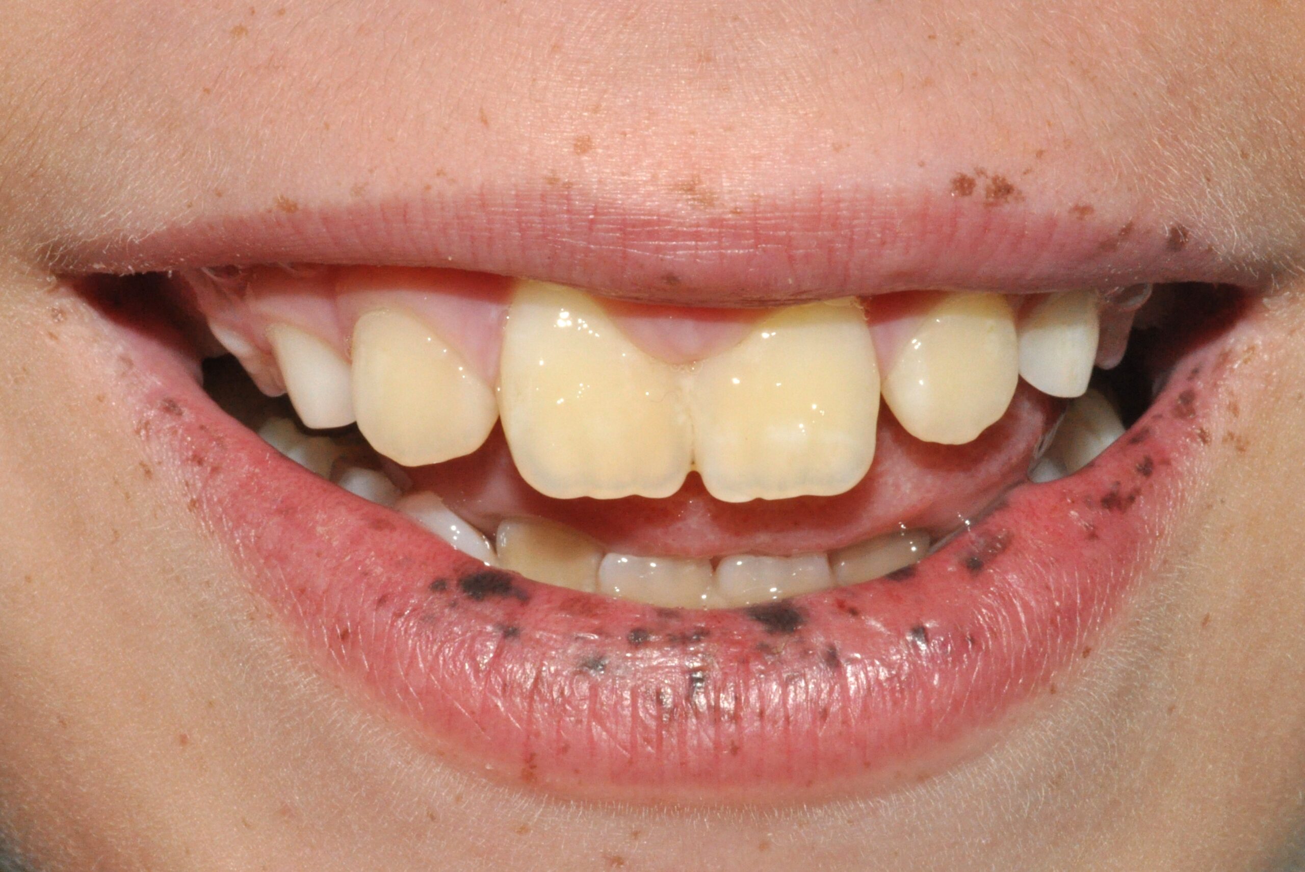
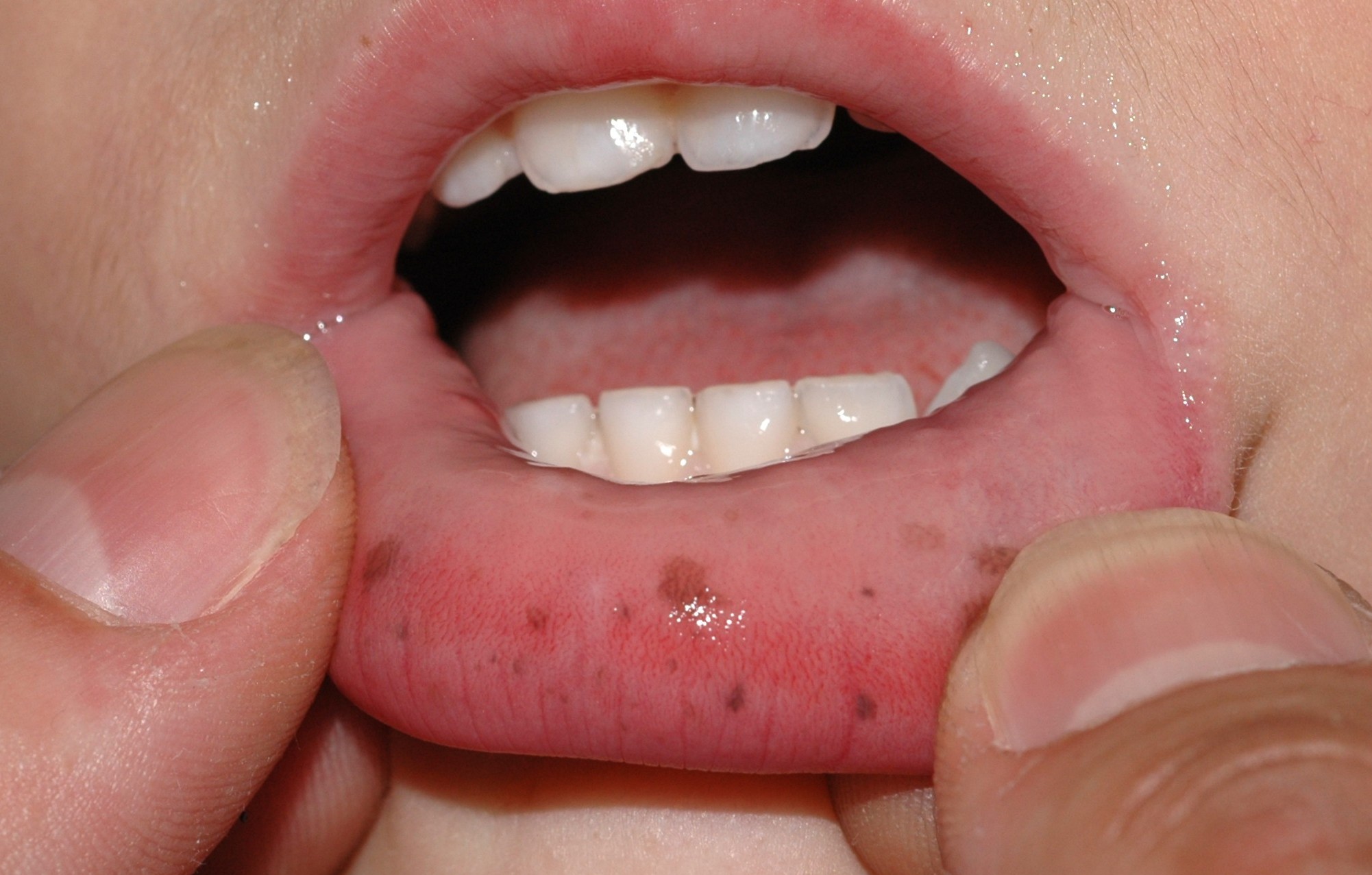
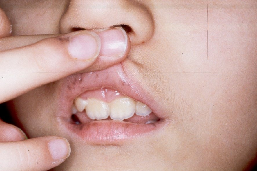
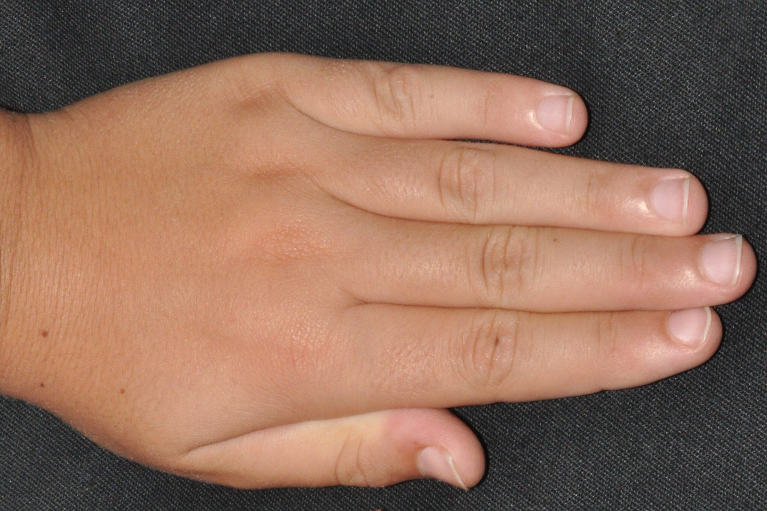
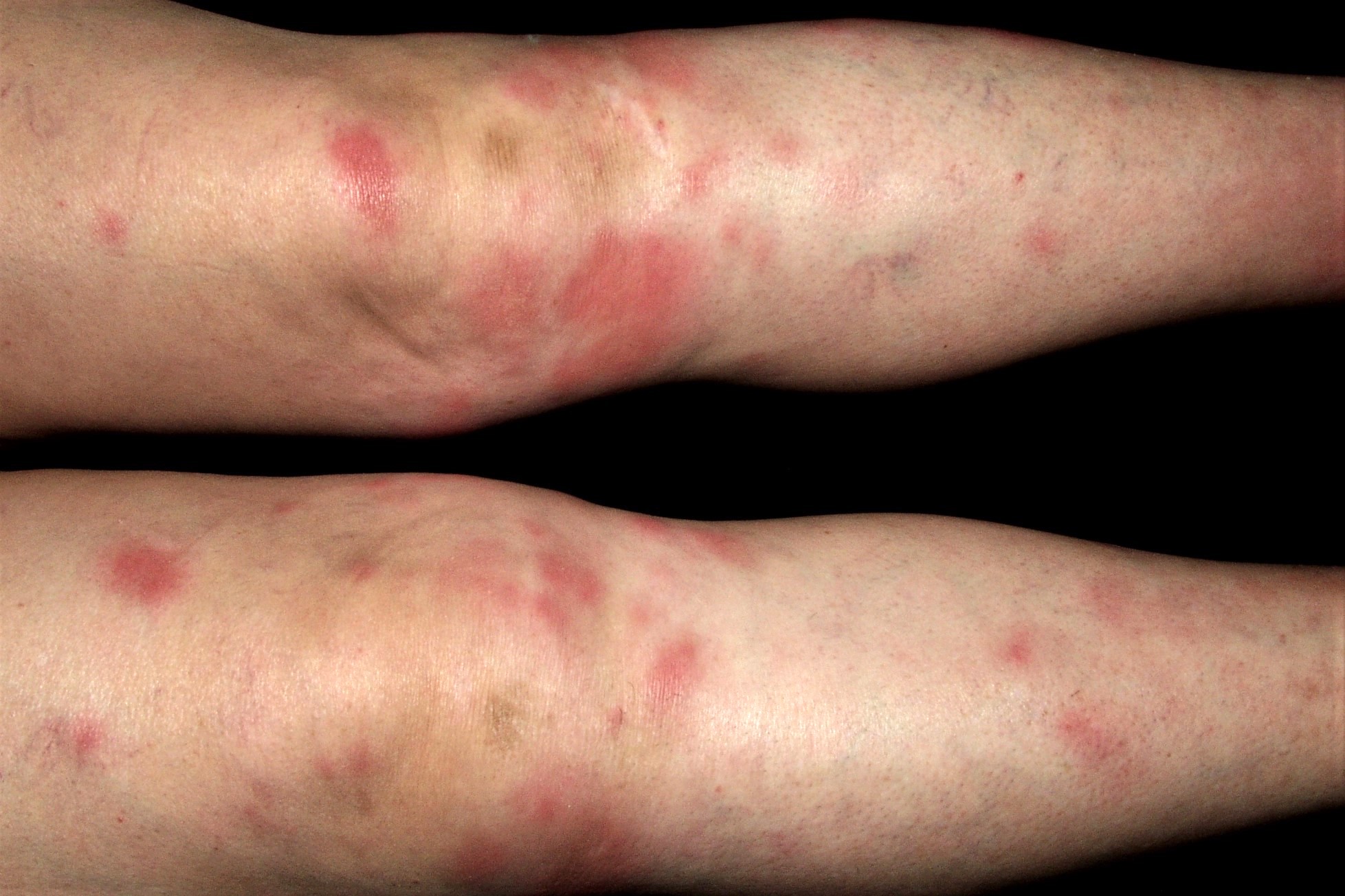
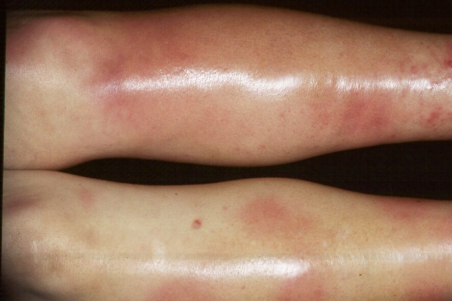
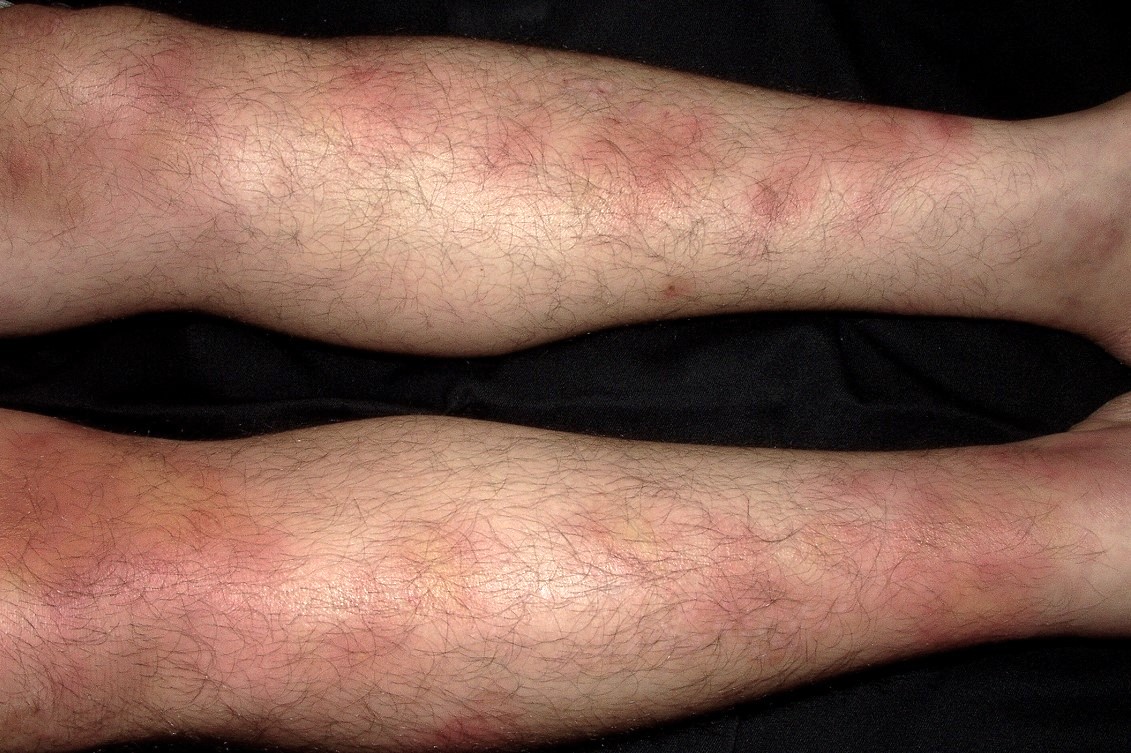
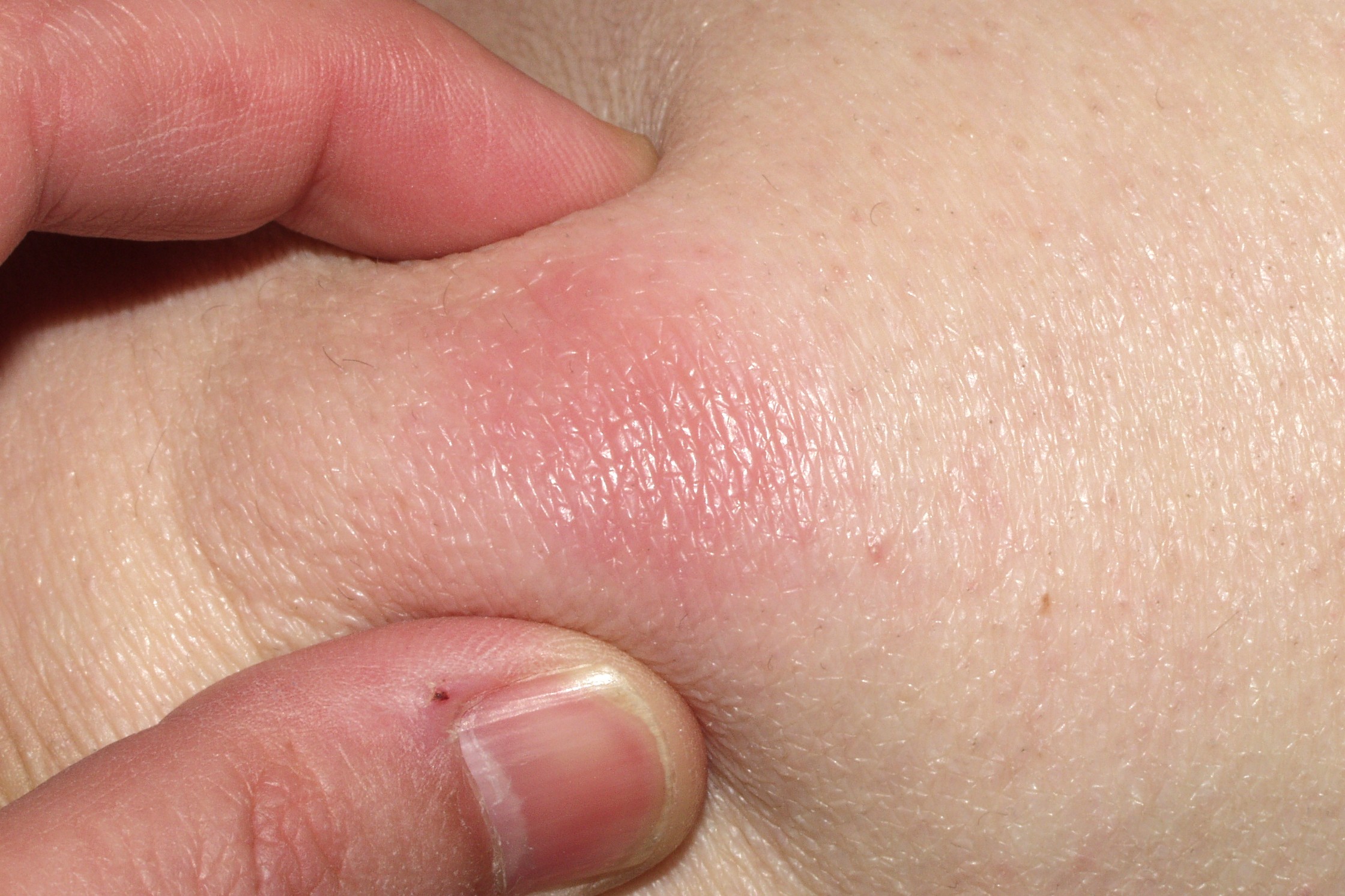
4. Bibliografía
-Contrò G, Tallerico R, Dattilo V, Fabiani F, Enzo MV, Hladnik U, Dastoli S, Nisticò SP, Colao E, Perrotti N, Iuliano R. A novel ABCC6 variant causative of pseudoxanthoma elasticum. Hum Genome Var. 2019; 20;6:30.
-Kim D, Seo EJ, Song YS, Suh CH, Kim JW, Kim DJ, Suh DC. Current Status of Clinical Diagnosis and Genetic Analysis of Hereditary Hemorrhagic Telangiectasia in South Korea: Multicenter Case Series and a Systematic Review. Neurointervention. 2019; 14:91-98.
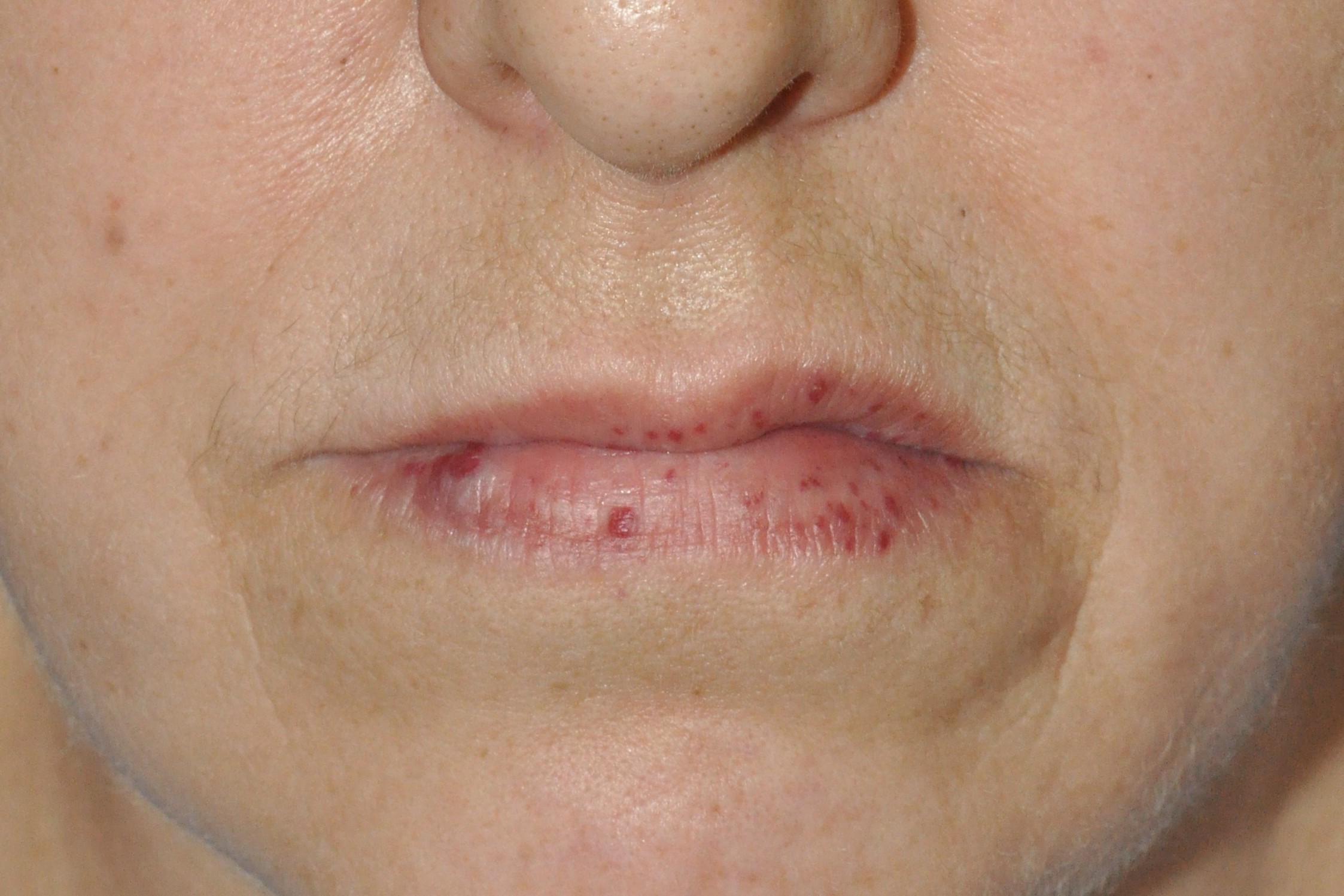
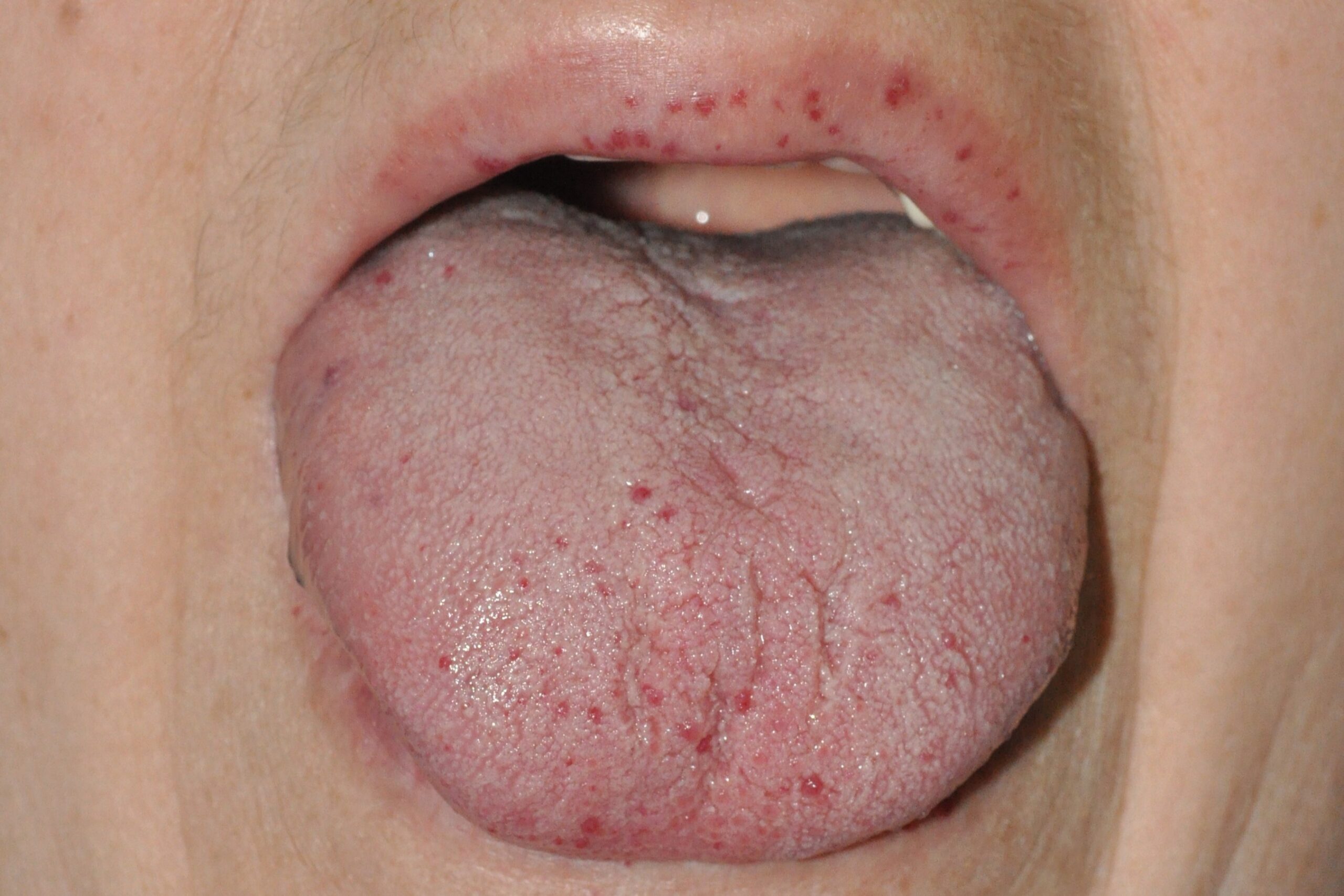
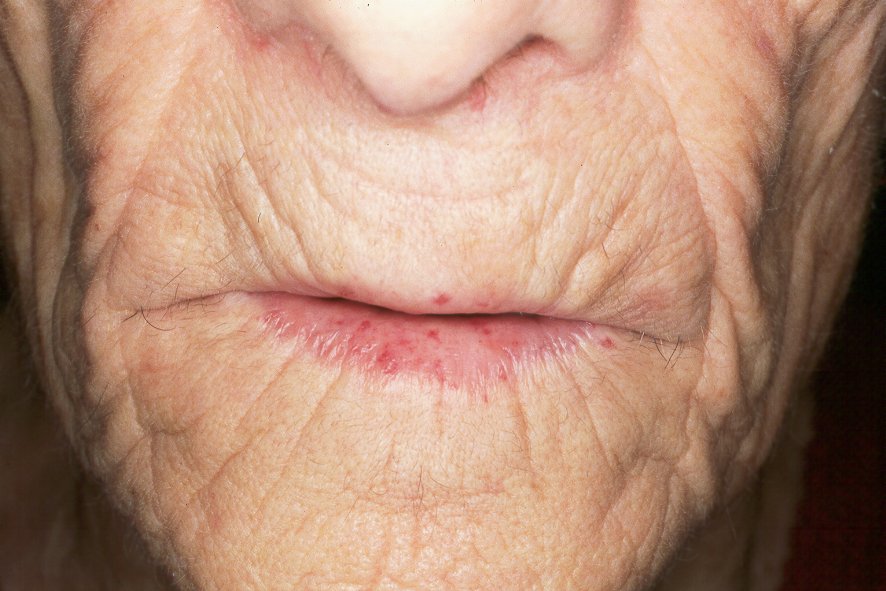
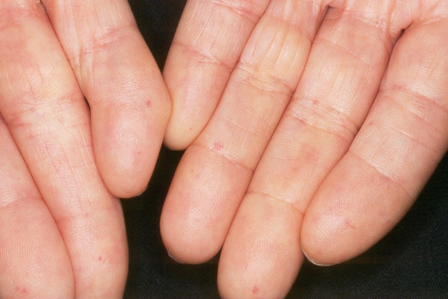
-Orizaga-Y-Quiroga TL, Villarreal-Martínez A, Jaramillo-Moreno G, Ocampo-Candiani J. Osler-Weber-Rendu Syndrome in Relation to Dermatology. Actas Dermosifiliogr. 2019; 110:526-532.
-Tortora A, Riccioni ME, Gaetani E, Ojetti V, Holleran G, Gasbarrini A. Rendu-Osler-Weber disease: a gastroenterologist’s perspective. Orphanet J Rare Dis. 2019; 14:130.
-Dupuis-Girod S, Cottin V, Shovlin CL. The Lung in Hereditary Hemorrhagic Telangiectasia. Respiration. 2017; 94:315-330.
-Robard L, Michel J, Prulière Escabasse V, Bequignon E, Vérillaud B, Malard O, Crampette L; SFORL Work-Group. Guidelines of the French Society of Otorhinolaryngology (SFORL) (short version). Specific treatment of epistaxis in Rendu-Osler-Weber disease. Eur Ann Otorhinolaryngol Head Neck Dis. 2017; 134:37-41.
-Contegiacomo A, Del Ciello A, Rella R, Attempati N, Coppolino D, Larici AR, Di Stasi C, Marano G, Manfredi R. Pulmonary arteriovenous malformations: what the interventional radiologist needs to know. Radiol Med. 2019. doi: 10.1007/s11547-019-01051-7.
-Pujol RM, Casanova JM, Egido R, Pujol J, de Moragas JM. Multiple familial pilomatricomas: a cutaneous marker for Gardner syndrome? Pediatr Dermatol. 1995; 12:331-5.
-Seehra J, Patel S, Bryant C. Gardner’s Syndrome revisited: a clinical case and overview of the literature. J Orthod. 2016; 43:59-64.
-Juhn E, Khachemoune A. Gardner syndrome: skin manifestations, differential diagnosis and management. Am J Clin Dermatol. 2010;11:117-22.
-Khanna K, Khanna V, Bhatnagar V. Peutz-Jeghers syndrome: need for early screening. BMJ Case Rep. 2018; 11. pii: e225076
-Wu M, Krishnamurthy K. Peutz-Jeghers Syndrome. En: StatPearls [Internet]. Treasure Island (FL): StatPearls Publishing; 2018-2019.
-Horikawa H, Hirai I, Umegaki-Arao N, Amagai M, Saito M. Alopecia in Cronkhite-Canada syndrome: Is it truly telogen effluvium? Australas J Dermatol. 2019; doi: 10.1111/ajd.13073.
-Li Y, Luo HQ, Wu D, Xue XW, Luo YF, Xiao YM, Chen J, Zhou WX. Clinicopathologic features of Cronkhite-Canada syndrome and the significance of IgG4-positive plasma cells infiltration. Zhonghua Bing Li Xue Za Zhi. 2018; 47:753-757.
-Richter L, Rappersberger K. Hauterscheinungen bei chronisch entzündlichen Darmerkrankungen: Morbus Crohn und Colitis ulcerosa. Hautarzt. 2016; 67:940-947.
-Cui RZ, Bruce AJ, Rogers RS 3rd. Recurrent aphthous stomatitis. Clin Dermatol. 2016; 34:475-81.
-Farhi D, Cosnes J, Zizi N, Chosidow O, Seksik P, Beaugerie L, Aractingi S, Khosrotehrani K. Significance of erythema nodosum and pyoderma gangrenosum in inflammatory bowel diseases: a cohort study of 2402 patients. Medicine (Baltimore). 2008; 87:281-93.
-Filosa A, Filosa G. Neutrophilic dermatoses: a broad spectrum of disease. G Ital Dermatol Venereol. 2018; 153:265-272.
-Kridin K, Cohen AD, Amber KT. Underlying Systemic Diseases in Pyoderma Gangrenosum: A Systematic Review and Meta-Analysis. Am J Clin Dermatol. 2018; 19:479-487.
-Plumptre I, Knabel D, Tomecki K. Pyoderma Gangrenosum: A Review for the Gastroenterologist. Inflamm Bowel Dis. 2018; 24:2510-2517.
-Maverakis E, Ma C, Shinkai K, Fiorentino D, Callen JP, Wollina U, Marzano AV, Wallach D, Kim K, Schadt C, Ormerod A, Fung MA, Steel A, Patel F, Qin R, Craig F, Williams HC, Powell F, Merleev A, Cheng MY. Diagnostic Criteria of Ulcerative Pyoderma Gangrenosum: A Delphi Consensus of International Experts. JAMA Dermatol. 2018; 154:461-466.
-Toh JWT, Young CJ, Rickard MJFX, Keshava A, Stewart P, Whiteley I. Peristomal pyoderma gangrenosum: 12-year experience in a single tertiary referral centre. ANZ J Surg. 2018; 88:E693-E697.
-Partridge ACR, Bai JW, Rosen CF, Walsh SR, Gulliver WP, Fleming P. Effectiveness of systemic treatments for pyoderma gangrenosum: a systematic review of observational studies and clinical trials. Br J Dermatol. 2018; 179:290-295.
-Cohen PR. Sweet’s síndrome -a comprehensive review of an acute febrile neutrophilic dermatosis. Orphanet J Rare Dis. 2007; 2:34.
–Villarreal-Villarreal CD, Ocampo-Candiani J, Villarreal-Martínez A. Sweet Syndrome: A Review and Update. Actas Dermosifiliogr. 2016; 107:369-78.
-Nelson CA, Noe MH, McMahon CM, Gowda A, Wu B, Ashchyan HJ, Perl AE, James WD, Micheletti RG, Rosenbach M. Sweet syndrome in patients with and without malignancy: A retrospective analysis of 83 patients from a tertiary academic referral center. J Am Acad Dermatol. 2018; 78:303-309.
-Nofal A, Abdelmaksoud A, Amer H, Nofal E, Yosef A, Gharib K, Albalat W, Eldesouky F, Ebrahim HM, Abdelshafy AS, Fayed H. Sweet’s syndrome: diagnostic criteria revisited. J Dtsch Dermatol Ges. 2017; 15:1081-1088.
-Cohen PR. Sweet’s síndrome -a comprehensive review of an acute febrile neutrophilic dermatosis. Orphanet J Rare Dis. 2007; 2:34.
-Raza S, Kirkland RS, Patel AA, Shortridge JR, Freter C. Insight into Sweet’s syndrome and associated-malignancy: a review of the current literature. Int J Oncol. 2013; 42:1516-22.
-Rochet NM, Chavan RN, Cappel MA, Wada DA, Gibson LE. Sweet syndrome: clinical presentation, associations, and response to treatment in 77 patients. J Am Acad Dermatol. 2013; 69:557-64.
-Nelson CA, Stephen S, Ashchyan HJ, James WD, Micheletti RG, Rosenbach M. Neutrophilic dermatoses: Pathogenesis, Sweet syndrome, neutrophilic eccrine hidradenitis, and Behçet disease. J Am Acad Dermatol. 2018; 79:987-1006.
-Raza S, Kirkland RS, Patel AA, Shortridge JR, Freter C. Insight into Sweet’s syndrome and associated-malignancy: a review of the current literature. Int J Oncol. 2013; 42:1516-22.
-Mullins TB, Zito PM. Reactive Perforating Collagenosis. StatPearls. Treasure Island (FL): StatPearls Publishing; 2019 Jan-2019 Aug 3.
-Marano AL, Selim MA, Cardones AR, Burton CS. Nodular amyloidosis in a patient with systemic scleroderma. Dermatol Online J. 2018; 24(9). pii: 13030/qt85b030dk.
-Yang M, Li LF, Chang JM. Histopathological findings of primary cutaneous lichenoid and macular amyloidosis. Zhongguo Yi Xue Ke Xue Yuan Xue Bao. 2009; 31:31-3.
–D’Aguanno V, Ralli M, Artico M, Russo FY, Scarpa A, Fiore M, Tirassa P, Severini C, de Vincentiis M, Greco A. Systemic Amyloidosis: a Contemporary Overview. Clin Rev Allergy Immunol. 2019. doi: 10.1007/s12016-019-08759-4.
-Hazenberg BP. Amyloidosis: a clinical overview. Rheum Dis Clin North Am. 2013; 39:323-45.
-Vyas K, Morgaonkar M, Gupta S, Jain SK. Primary Systemic Amyloidosis with Unusual Dermatological Manifestations: A Rare Case Report. Indian J Dermatol. 2016; 61:216-8.
-Kumar S, Sengupta RS, Kakkar N, Sharma A, Singh S, Varma S. Skin involvement in primary systemic amyloidosis. Mediterr J Hematol Infect Dis. 2013; 5:e2013005.
-Flores-Bozo LR, Echevarría-Keel J, Domínguez-Cherit J, Esquivel-Pedraza L, Méndez-Flores S. Mucocutaneous manifestations in systemic amyloidosis A retrospective analytical study in a tertiary care center. Int J Dermatol. 2019; 58:1062-1068.
-Wechalekar AD, Gillmore JD, Hawkins PN. Systemic amyloidosis. Lancet. 2016; 387(10038):2641-2654.
–Weidner T, Illing T, Elsner P. Primary Localized Cutaneous Amyloidosis: A Systematic Treatment Review. Am J Clin Dermatol. 2017; 18:629-642.
-Choi JY, Sippe J, Lee S. Acitretin for lichen amyloidosus. Australas J Dermatol. 2008; 49:109-13.
-Grimmer J, Weiss T, Weber L, Meixner D, Scharffetter-Kochanek K. Successful treatment of lichen amyloidosis with combined bath PUVA photochemotherapy and oral acitretin. Clin Exp Dermatol. 2007; 32:39-42.
-García-Lozano JA, Ocampo-Candiani J, Martínez-Cabriales SA, Garza-Rodríguez V. An Update on Calciphylaxis. Am J Clin Dermatol. 2018; 19:599-608.
-Jiménez-Gallo D, Ossorio-García L, Linares-Barrios M. Calcinosis Cutis and Calciphylaxis. Actas Dermosifiliogr. 2015; 106:785-94; Cucchiari D, 2018.
-Cucchiari D, Torregrosa JV. Calciphylaxis in patients with chronic kidney disease: A disease which is still bewildering and potentially fatal. Nefrología. 2018; 38:579-586.
-Nigwekar SU. Calciphylaxis. Curr Opin Nephrol Hypertens. 2017; 26:276-281.
-Dutta P, Chaudet KM, Nazarian RM, Kroshinsky D, Nigwekar SU. Correlation between clinical and pathological features of cutaneous calciphylaxis. PLoS One. 2019; 14:e0218155.
-Yu WY, Bhutani T, Kornik R, Pincus LB, Mauro T, Rosenblum MD, Fox LP. Warfarin-Associated Nonuremic Calciphylaxis. JAMA Dermatol. 2017; 153:309-314.
–Galassi A, Perna F, De Nicola E, Moneghini L, Sganzaroli AB, Cozzolino M. Calciphylaxis in a dialysis patient treated by intralesional and systemic sodium thiosulphate on top of multifactorial intervention. Clin Kidney J. 2018; 12:546-549.
-Zuhaili B, Al-Talib K. Successful Treatment of Single Infected Calciphylaxis Lesion With Intralesional Injection of Sodium Thiosulfate at High Concentration. Wounds. 2019; 31:E54-E57.
-Murphy-Chutorian B, Han G, Cohen SR. Dermatologic manifestations of diabetes mellitus: a review. Endocrinol Metab Clin North Am. 2013; 42:869-98.
–Sibbald C, Reid S, Alavi A. Necrobiosis Lipoidica. Dermatol Clin. 2015; 33:343-60.
–Grillo E, Rodriguez-Muñoz D, González-Garcia A, Jaén P. Necrobiosis lipoidica. Aust Fam Physician. 2014; 43:129-30.
-Matas-Nadal C, Fernández-Armenteros JM, Casanova Seuma JM, Vilardell F, Baradad M, Aguayo-Ortiz RS. Papular spreading necrobiosis lipoidica: a rare clinical presentation of this pathology. Clin Exp Dermatol. 2019; 44:e159-e160.
-Hammer E, Lilienthal E, Hofer SE, Schulz S, Bollow E, Holl RW; DPV Initiative and the German BMBF Competence Network for Diabetes Mellitus. Risk factors for necrobiosis lipoidica in Type 1 diabetes mellitus. Diabet Med. 2017; 34:86-92.
-Peckruhn M, Tittelbach J, Elsner P. Update: Treatment of necrobiosis lipoidica. J Dtsch Dermatol Ges. 2017; 15:151-157.
-Lim JZ, Ng NS, Thomas C. Prevention and treatment of diabetic foot ulcers. J R Soc Med. 2017; 110:104-109).
-Zhang P, Lu J, Jing Y, Tang S, Zhu D, Bi Y. Global epidemiology of diabetic foot ulceration: a systematic review and meta-analysis †. Ann Med. 2017; 49:106-116.
-Blume P, Wu S. Updating the Diabetic Foot Treatment Algorithm: Recommendations on Treatment Using Advanced Medicine and Therapies. Wounds. 2018; 30:29-35.
-Bandyk DF. The diabetic foot: Pathophysiology, evaluation, and treatment. Semin Vasc Surg. 2018; 31:43-48.
-Noor S, Zubair M, Ahmad J. Diabetic foot ulcer–A review on pathophysiology, classification and microbial etiology. Diabetes Metab Syndr. 2015; 9:192-9.
-Ramirez-Acuña JM, Cardenas-Cadena SA, Marquez-Salas PA, Garza-Veloz I, Perez-Favila A, Cid-Baez MA, Flores-Morales V, Martinez-Fierro ML. Diabetic Foot Ulcers: Current Advances in Antimicrobial Therapies and Emerging Treatments. Antibiotics (Basel). 2019; 8. pii: E1939.
-Spanos K, Saleptsis V, Athanasoulas A, Karathanos C, Bargiota A, Chan P, Giannoukas AD. Factors Associated With Ulcer Healing and Quality of Life in Patients With diabetic Foot Ulcer. Angiology. 2017; 68:242-250.
–Fatourechi V. Pretibial myxedema: pathophysiology and treatment options. Am J Clin Dermatol. 2005; 6:295-309.
-Sabanova EA, Fadeyev VV, Potekaev NN, Lvov AN. Pretibial myxedema: pathogenetic features and clinical aspects. Probl Endokrinol (Mosk). 2019; 65:134-138.
-Anagnostis P, Artzouchaltzi A, Grekou A, Paschou SA, Gerou S, Tzalokostas V. Pretibial myxedema in a euthyroid patient. Hormones (Athens). 2018; 17:133-135.
-Zhang F, Lin XY, Chen J, Peng SQ, Shan ZY, Teng WP, Yu XH. Intralesional and topical glucocorticoids for pretibial myxedema: A case report and review of literature. World J Clin Cases. 2018; 6:854-861.
-Muray S, Casanova JM, Marco MP, Craver L, Martín M, Fernández E. Porfiria cutanea tarda, hemodialisis y hepatopatia VHC. Nefrologia. 2002; 22:570-3) se debe realizar un tratamiento combinado específico.
-de Schaetzen V, Richert B, de la Brassinne M. Les xanthomes. Rev Med Liege. 2004; 59:46-50.
-Zak A, Zeman M, Slaby A, Vecka M. Xanthomas: clinical and pathophysiological relations. Biomed Pap Med Fac Univ Palacky Olomouc Czech Repub. 2014; 158:181-8.
-Santiago L, Pinho A, Cardoso JC. Xantomas Eruptivos: Manifestação Cardinal de Distúrbio Metabólico Grave. Acta Med Port. 2018; 31:219-222.
-Duzayak S, Sayiner ZA, Erkılıç S, Inaloz HS. Acute pancreatitis with eruptive xanthoma. BMJ Case Rep. 2017; pii: bcr-2017-221543.
-Kong MX, Zhang Q, Cao L, Zhao C, Ru GQ, Bi Q. Familial hypercholesterolaemia with tuberous and tendinous xanthomas: case report and mutation analysis. Clin Exp Dermatol. 2015; 40:765-9.
-Koopal C, Visseren FL, Marais AD, Westerink J, Spiering W. Tendon xanthomas: Not always familial hypercholesterolemia. J Clin Lipidol. 2016; 10:1262-5.
-Zak A, Zeman M, Slaby A, Vecka M. Xanthomas: clinical and pathophysiological relations. Biomed Pap Med Fac Univ Palacky Olomouc Czech Repub. 2014; 158:181-8.
-Miyashiro D, Sanches JA. Paraneoplastic skin disorders: a review. G Ital Dermatol Venereol. 2016; 151:55-76.
-Gießen-Jung C, Wollenberg A, Reinholz M. [Paraneoplasms of the skin]. Internist (Berl). 2018; 59:134-144.
-Geng G, Yu X, Jiang J, Yu X. Aetiology and pathogenesis of paraneoplastic autoimmune disorders. Autoimmun Rev. 2019: 102422.
-Wick MR, Patterson JW. Cutaneous paraneoplastic syndromes. Semin Diagn Pathol. 2019; 36:211-228.
–Cheng E, Roy DB, Magro CM. A case of acanthosis nigricans coexisting with mycosis fungoides.Dermatol Online J. 2015; 21. pii: 13030/qt3sw2t3b8.
-Roser A, Pajot C, Kettani S, Michalak S, Le Clech C. Acanthosis nigricans malin révélant un cholangiocarcinome. Rev Med Interne. 2019; 40:750-753.
–Nguyen S, Grosber M, Gutermuth J. Acrokeratosis Can Be a Warning Sign of an Underlying Malignancy. Eur J Case Rep Intern Med. 2019; 6:001108.
-Räßler F, Goetze S, Elsner P. Acrokeratosis paraneoplastica (Bazex syndrome) – a systematic review on risk factors, diagnosis, prognosis and management.J Eur Acad Dermatol Venereol. 2017; 31:1119-1136.
-Gül Ü. Acral manifestations of paraneoplastic and collagen vascular diseases. Clin Dermatol. 2017; 35:50-5.
-Rao AG, Farheen SS, Amit K, Reddy UD, Aparna K, Kranthi J, Hukkani R. Erythema Gyratum Repens Associated With Diffuse B-cell Lymphoma-Report of A Rare Case. Indian J Dermatol. 2019; 64:338.
-Clyatt G, Cole J. A Unique Case of Localized Palmar Erythema Annulare Centrifugum. Cureus. 2022; 14(6):e25718.
-V’lckova-Laskoska M, Balabanova-Stefanova M, Arsovska-Bezhoska I, Caca-Biljanovska N, Laskoski D. Necrolytic Migratory Erythema: Complete Healing after Surgical Removal of Pancreatic Carcinoma. Acta Dermatovenerol Croat. 2018; 26:329-332.
-Wyatt JP, Anderson HF, Greer KE, Cordoro KM. Acquired hypertrichosis lanuginosa as a presenting sign of metastatic prostate cancer with rapid resolution after treatment. J Am Acad Dermatol. 2007; 56(2 Suppl):S45-7.
-Paolino G, Didona D, Magliulo G, Iannella G, Didona B, Mercuri SR, Moliterni E, Donati M, Ciofalo A, Granata G, Ranuzzi P, Falasca V, Calvieri S. Paraneoplastic Pemphigus: Insight into the Autoimmune Pathogenesis, Clinical Features and Therapy. Int J Mol Sci. 2017; 18. pii: E2532.
–Tirado-Sánchez A, Bonifaz A. Paraneoplastic Pemphigus. A Life-Threatening Autoimmune Blistering Disease. Actas Dermosifiliogr. 2017; 108:902-910.
-Yong AA, Tey HL. Paraneoplastic pemphigus. Australas J Dermatol. 2013; 54:241-50.
-Maruta CW, Miyamoto D, Aoki V, Carvalho RGR, Cunha BM, Santi CG. Paraneoplastic pemphigus: a clinical, laboratorial, and therapeutic overview. An Bras Dermatol. 2019; 94:388-398.
–Stoyanov GS, Dzhenkov DL, Tzaneva M. Thrombophlebitis Migrans (Trousseau Syndrome) in Pancreatic Adenocarcinoma: An Autopsy Report. Cureus. 2019; 11:e5528.
–Cunha DG, Campos-do-Carmo G, Marujo JM, Verardino GC. Paraneoplastic Sweet’s syndrome. An Bras Dermatol. 2018; 93:576-578.
-Martires K, Callahan S, Terushkin V, Brinster N, Leger M, Soter NA. Eosinophilic dermatosis of hematologic malignancy. Dermatol Online J. 2016; 22. pii: 13030/qt8467m0j9.
-Weisshaar E, Weiss M, Mettang T, Yosipovitch G, Zylicz Z; Special Interest Group of the International Forum on the Study of Itch. Paraneoplastic itch: an expert position statement from the Special Interest Group (SIG) of the International Forum on the Study of Itch (IFSI). Acta Derm Venereol. 2015; 95:261-5.
-Rowe B, Yosipovitch G. Paraneoplastic Itch Management. Curr Probl Dermatol. 2016; 50:149-54.
–Laidler NK. Dermatomyositis as a paraneoplastic phenomenon in oesophageal cancer. BMJ Case Rep. 2018; 11.
-Aguayo-Ortiz R, Casanova-Seuma JM. Dermatomiositis amiopática. Piel: formación continuada en dermatología, 2012; 27:77-81.
-Shah AA, Casciola-Rosen L. Cancer and scleroderma: a paraneoplastic disease with implications for malignancy screening. Curr Opin Rheumatol. 2015; 27:563-70.
-Santos-Faria D, Leite Silva J, Sousa-Neves J, Ramos Rodrigues J, Afonso C, Peixoto D. Systemic sclerosis and myositis as a paraneoplastic syndrome secondary to multiple myeloma. Acta Reumatol Port. 2018; 43:316-317.
-Alcaraz I, Cerroni L, Rütten A, Kutzner H, Requena L. Cutaneous metastases from internal malignancies: a clinicopathologic and immunohistochemical review. Am J Dermatopathol. 2012; 34:347-93.
-Strickley JD, Jenson AB, Jung JY. Cutaneous Metastasis. Hematol Oncol Clin North Am. 2019; 33:173-197.
-Hussein MR. Skin metastasis: a pathologist’s perspective. J Cutan Pathol. 2010; 37:e1-20.
-Hager CM, Cohen PR. Cutaneous lesions of metastatic visceral malignancy mimicking pyogenic granuloma. Cancer Invest. 1999; 17:385-90.
-Otsuka I. Cutaneous Metastases in Ovarian Cancer. Cancers (Basel). 2019; 11. pii: E1292.
-Vila Justribó M, Casanova Seuma JM, Portero L, Egido R. Metástasis cutáneas como primera manifestación de carcinoma broncogénico. Arch Bronconeumol 1994; 30: 314-6.
-Mollet TW, Garcia CA, Koester G. Skin metastases from lung cancer. Dermatol Online J. 2009; 15:1.
–Feeney KJ, Mastrangelo MJ. Intralesional therapy for in-transit and satellite metastases in melanoma. Surg Oncol Clin N Am. 2015; 24:299-308.
-Adams SJ, Kanthan R. Paget’s disease of the male breast in the 21st century: A systematic review. Breast. 2016; 29:14-23.
– Sandoval-Leon AC, Drews-Elger K, Gomez-Fernandez CR, Yepes MM, Lippman ME. Paget’s disease of the nipple. Breast Cancer Res Treat. 2013; 141:1-12.
-Moore SA, Notgrass HM, Vandergriff TW, Sahoo S. Mammary Paget’s Disease of the Male Breast: A Rare Case With an Unusual Immunohistochemical Profile. Int J Surg Pathol. 2019:1066896919874878
-Drews-Elger K, Sandoval-Leon AC, Ergonul AB, Jegg AM, Gomez-Fernandez C, Miller PC, El-Ashry D, Lippman ME. Paget’s disease of the nipple in a Her2-positive breast cancer xenograft model. Breast Cancer Res Treat. 2019. doi: 10.1007/s10549-019-05490-8.
-Wei Y, Zhu Q, Li J, Jiang Y. Clinical and Sonographic Features of Mammary Paget’s Disease. Zhongguo Yi Xue Ke Xue Yuan Xue Bao. 2017; 39:396-400.
-Geffroy D, Doutriaux-Dumoulins I, Labbe-Devilliers C, Meingan P, Houdebine S, Sagan C, Dejode M, Ricaud-Couprie M. Maladie de Paget du mamelon et principaux diagnostics différentiels. J Radiol. 2011; 92:889-98.
-Hudson-Phillips S, Cox K, Patel P, Al Sarakbi W. Paget’s disease of the breast: diagnosis and management. Br J Hosp Med (Lond). 2023; 84(1):1-8.
-Fernández-Figueras MT, Puig L, Casanova JM, Musulén E, Matías-Guiu X, Navas-Palacios JJ. Pigmented epidermotropic ductal carcinoma of the breast in a male. Ultrastructural evidence of melanocytic colonization and melanin transfer to the tumor. J Cutan Pathol. 1995; 22:276-80.
-Lopes Filho LL, Lopes IM, Lopes LR, Enokihara MM, Michalany AO, Matsunaga N. Mammary and extramammary Paget’s disease. An Bras Dermatol. 2015; 90:225-31.
–Primo WQSP, Primo GRP, Basilio DB, Machado KK, Carvalho JP, Carvalho FM. Vulvar Paget disease secondary to high-grade urothelial carcinoma with underlying massive vascular embolization and cervical involvement: case report of unusual presentation. Diagn Pathol. 2019; 14:125.
-Nasioudis D, Bhadra M, Ko EM. Extramammary Paget disease of the vulva: Management and prognosis. Gynecol Oncol. 2019. pii: S0090-8258(19)31638-5.
-Mammino JR, Daze RP, Mammino J. Fungal Masquerader: A Case Report of Extramammary Paget’s Disease Treated with Radiotherapy. Cureus. 2019;11:e5937.