
Currículum
Tema 09. Enfermedades vesiculosas y ampollosas
1. INTRODUCCIÓN
0/1

2. ENFERMEDADES AMPOLLOSAS AGUDAS
0/2
3. DERMATOSIS AMPOLLOSAS CRÓNICAS AUTOINMUNES
0/4
4. DERMATOSIS AMPOLLOSAS CRÓNICAS HEREDITARIAS
0/2
5. BIBLIOGRAFÍA
0/1
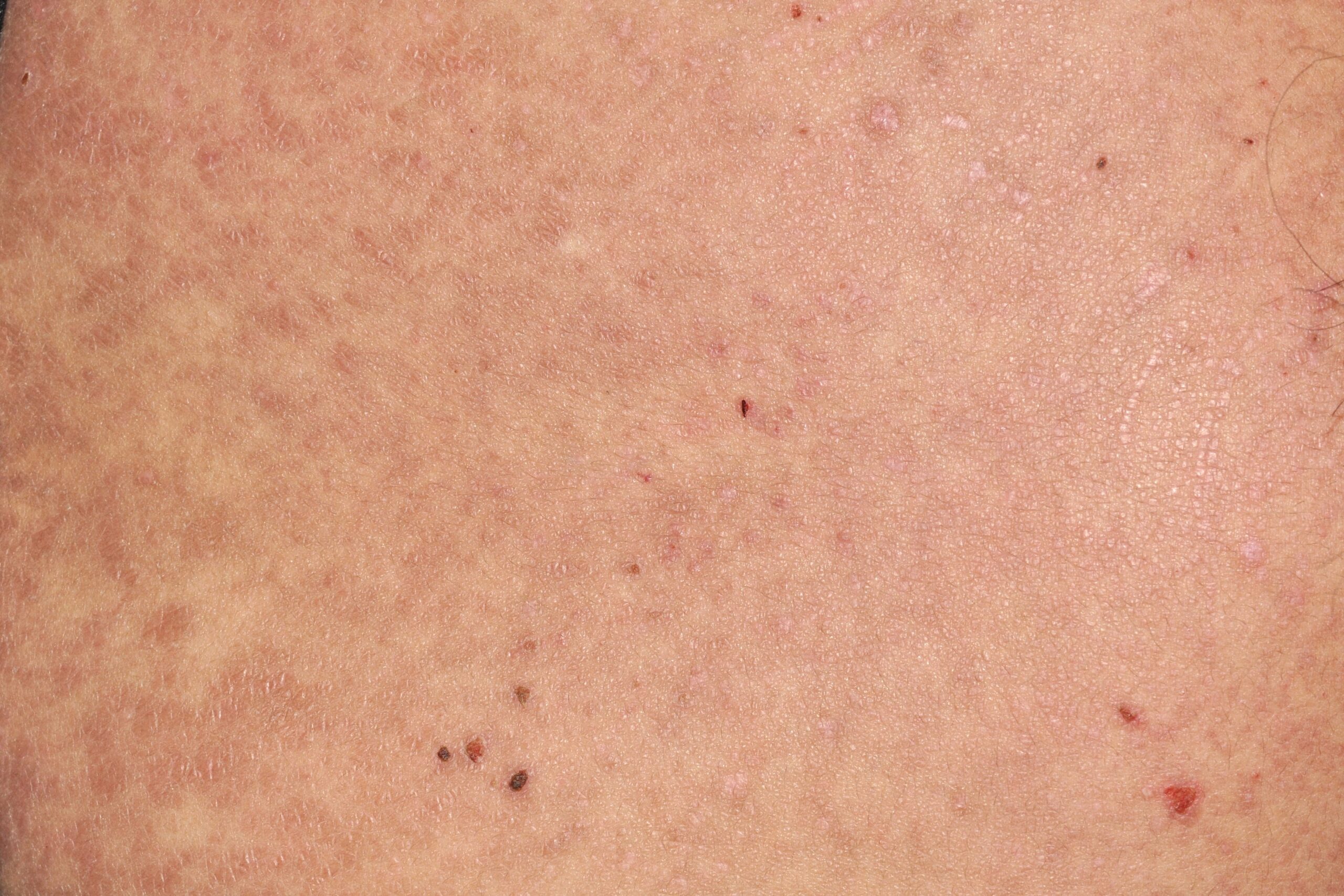
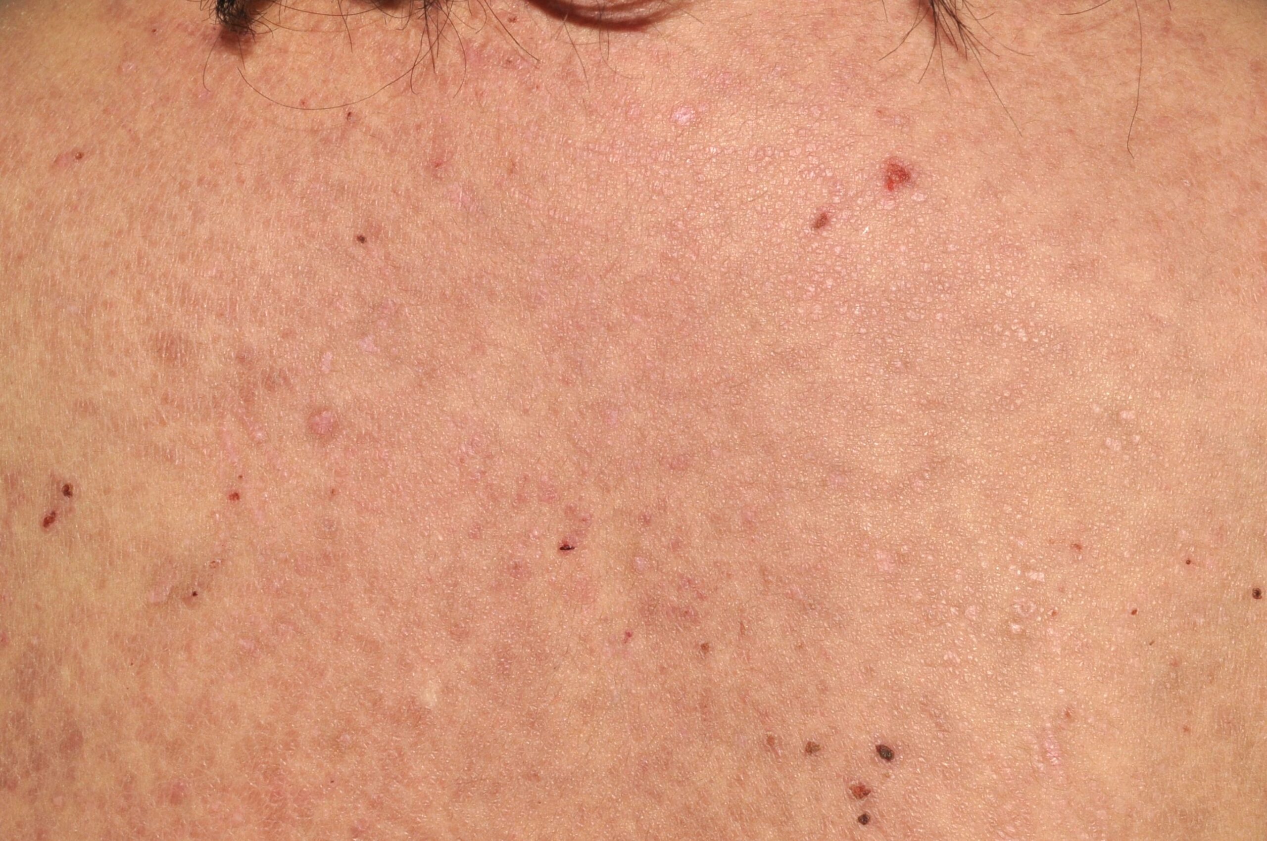
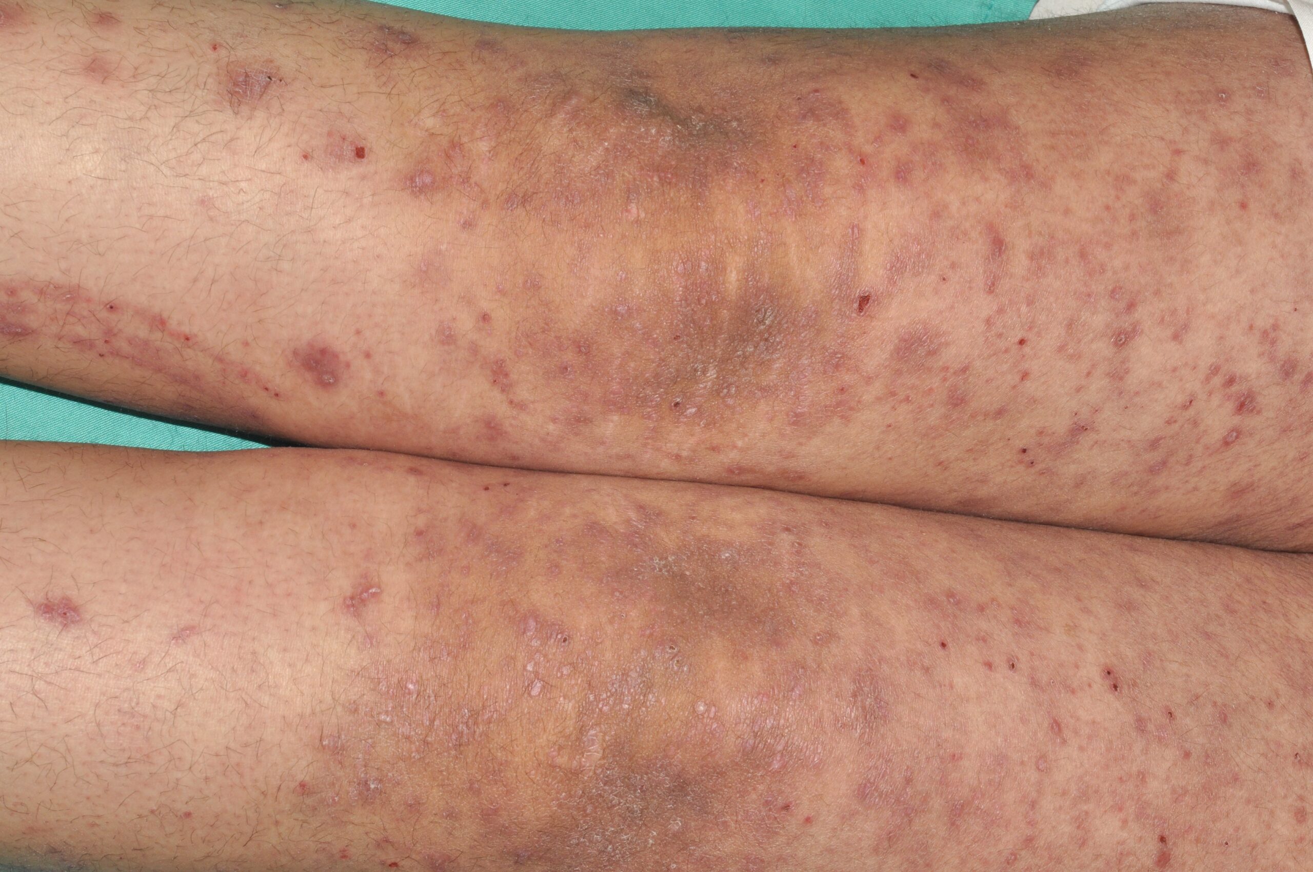
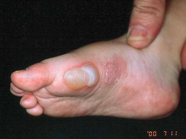
Imágenes tema

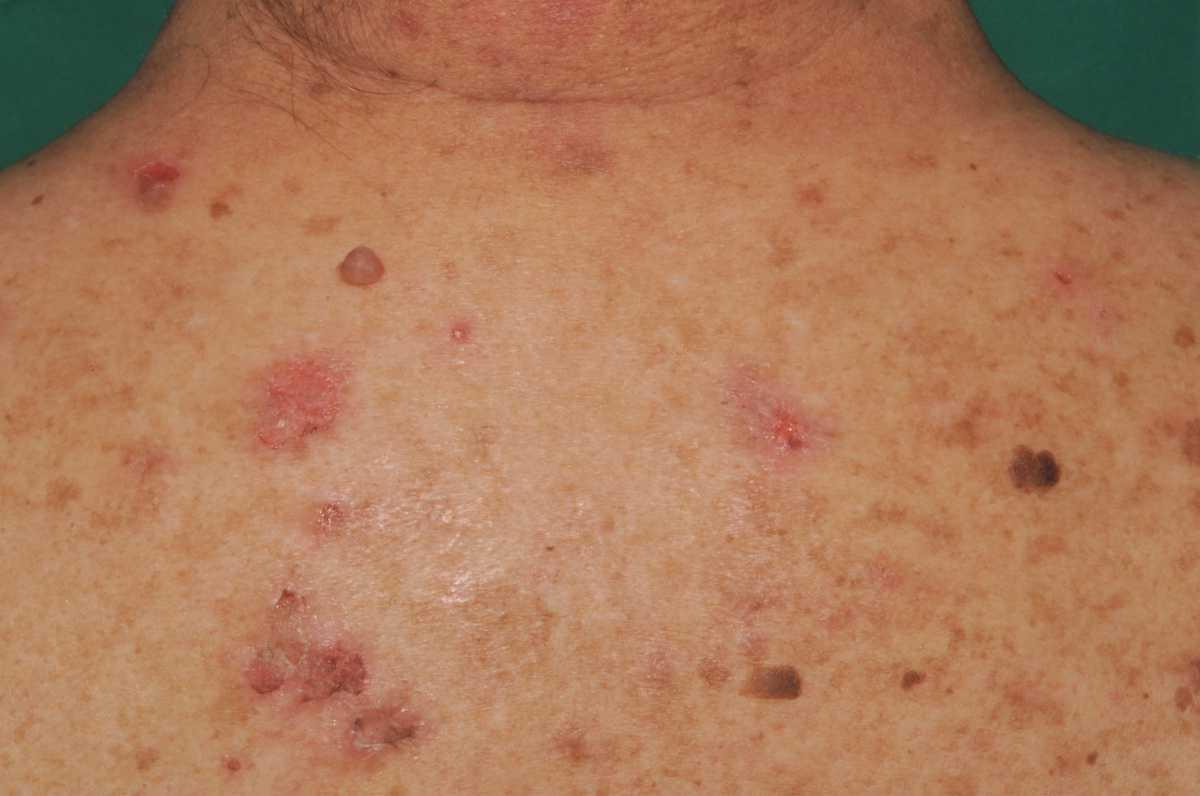
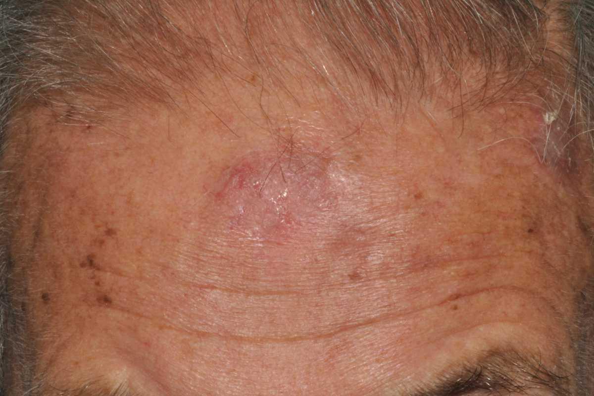
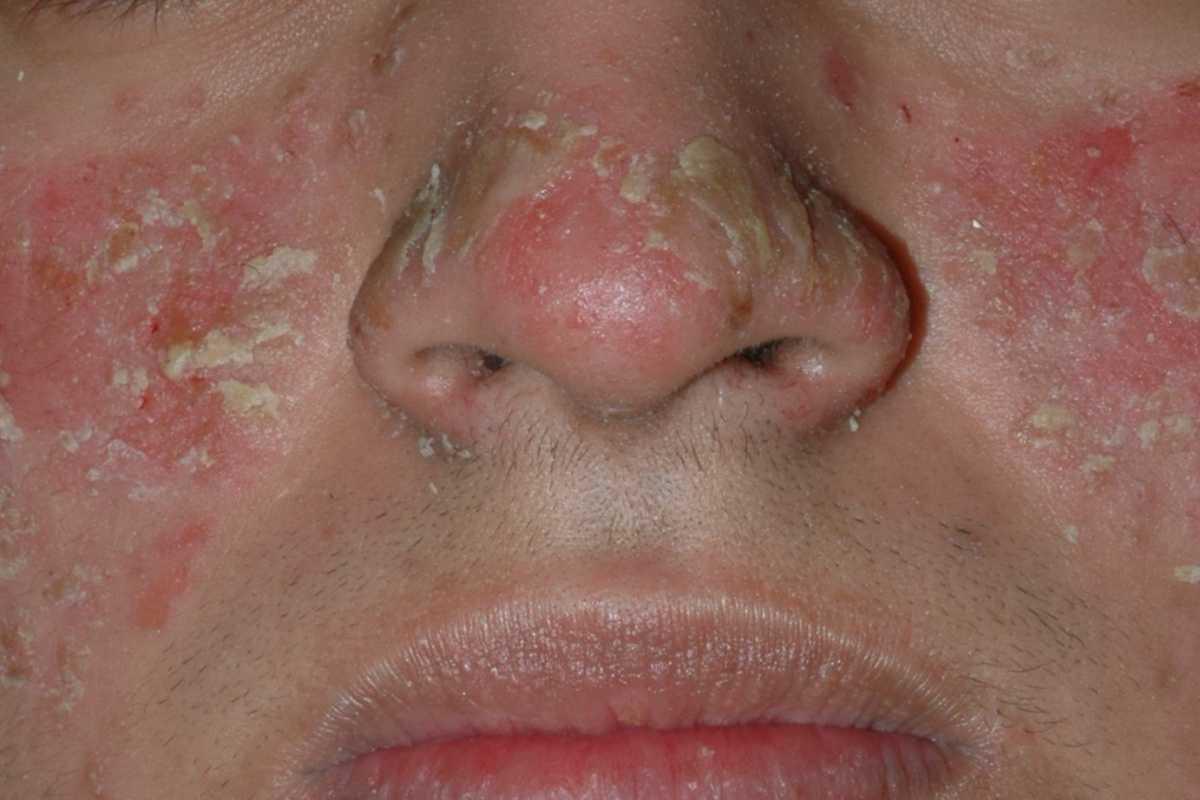
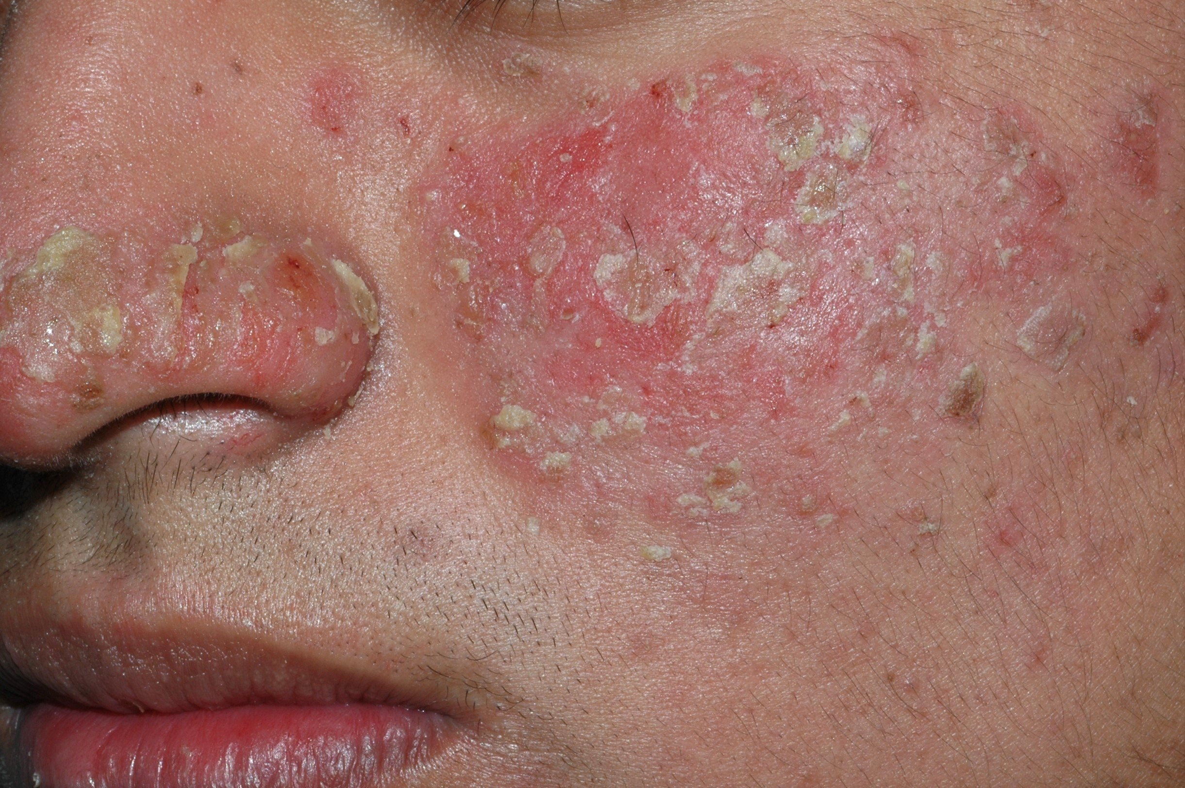
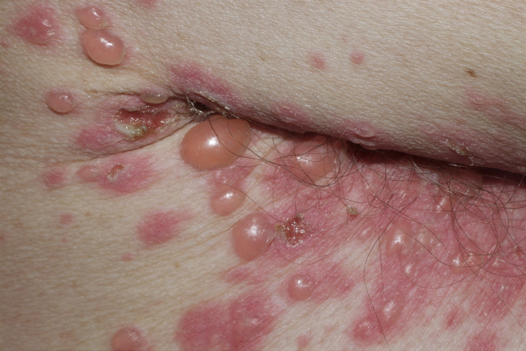
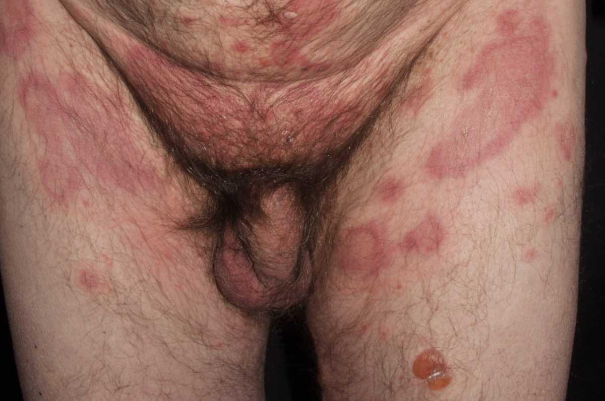
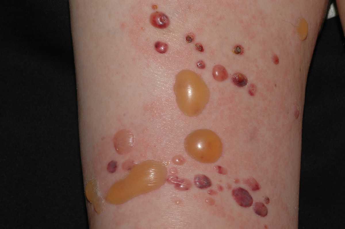
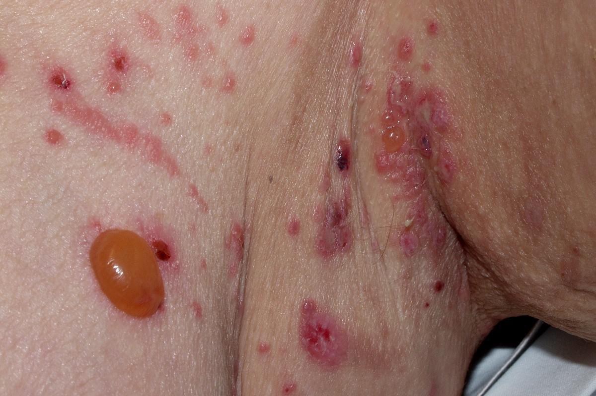
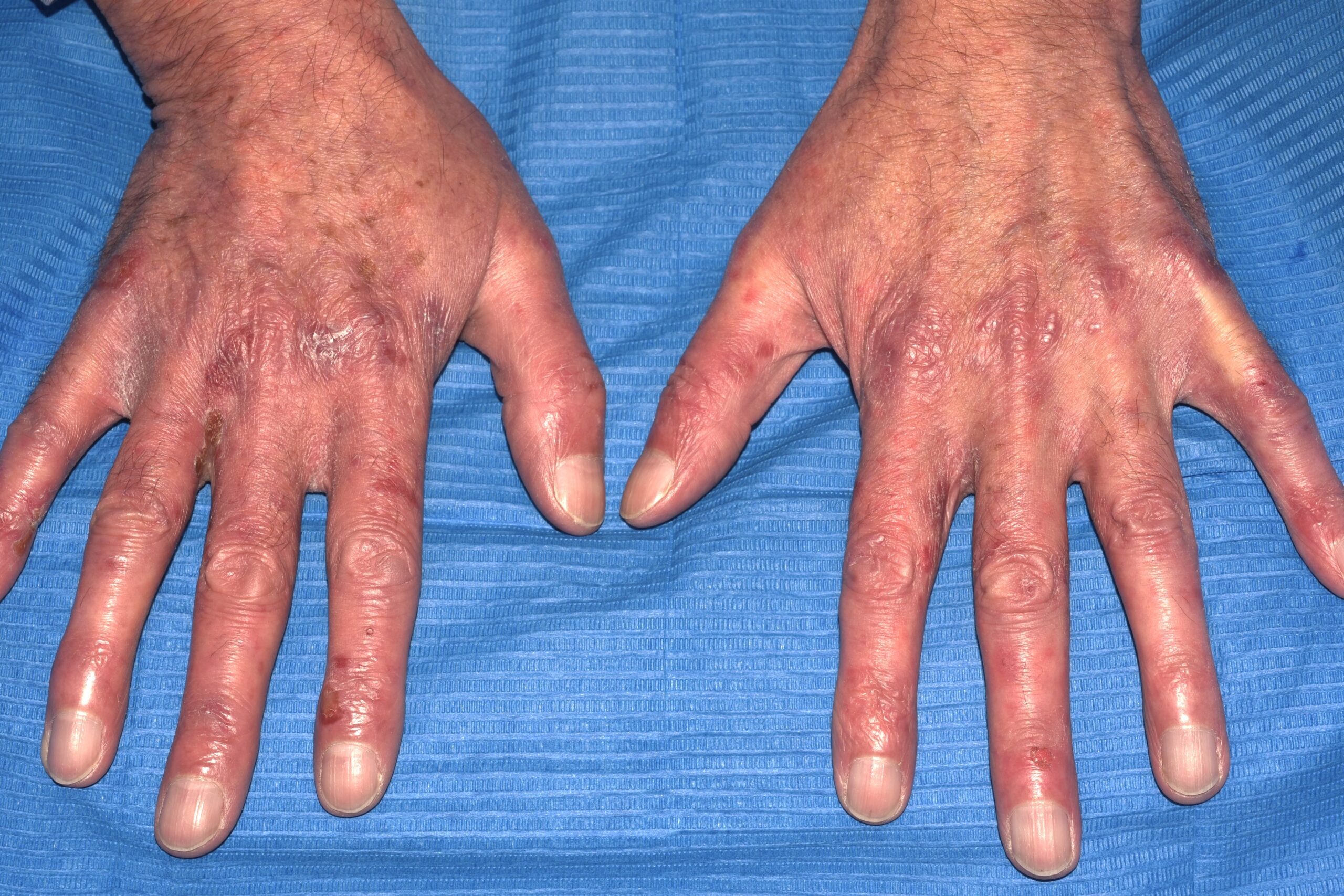
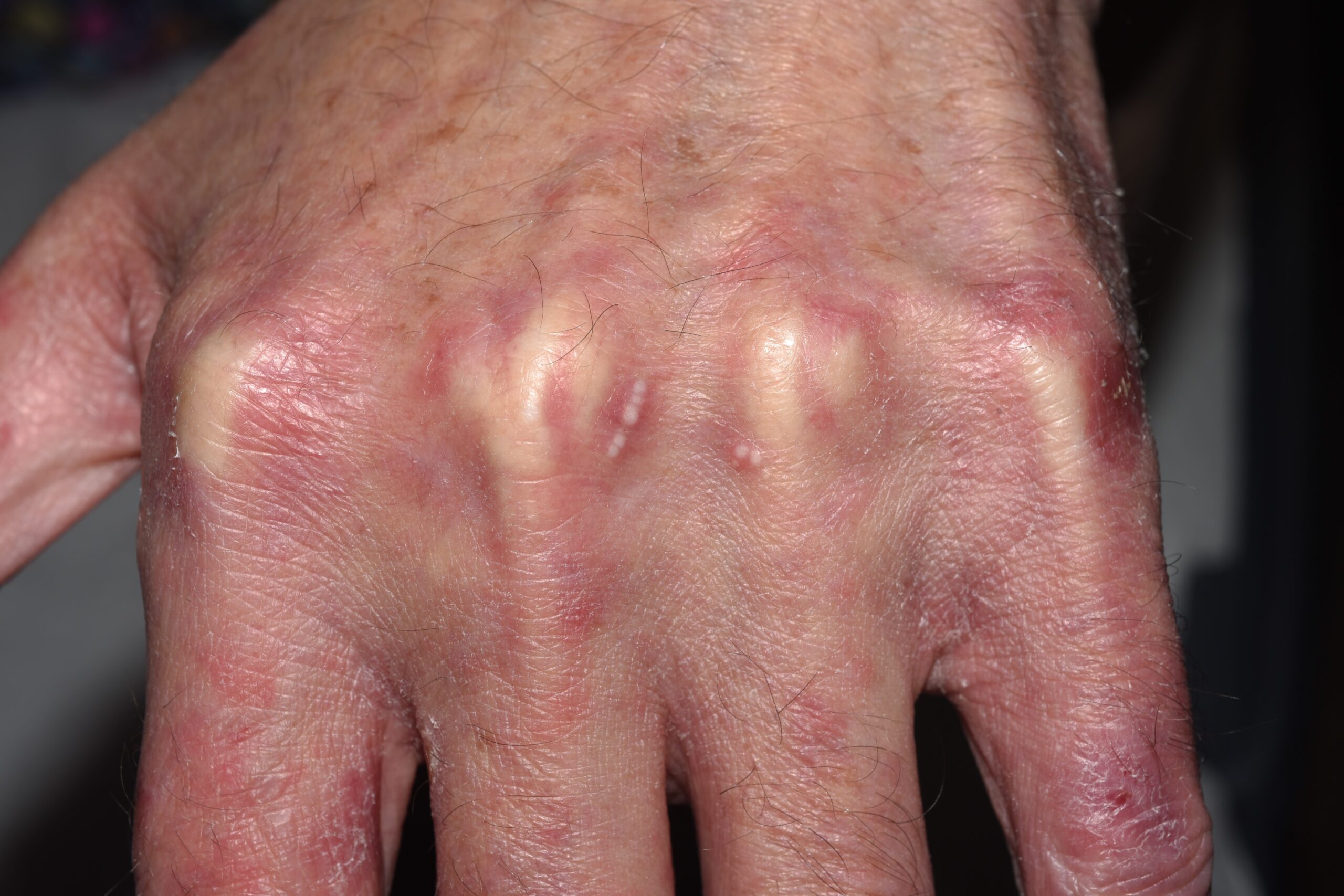
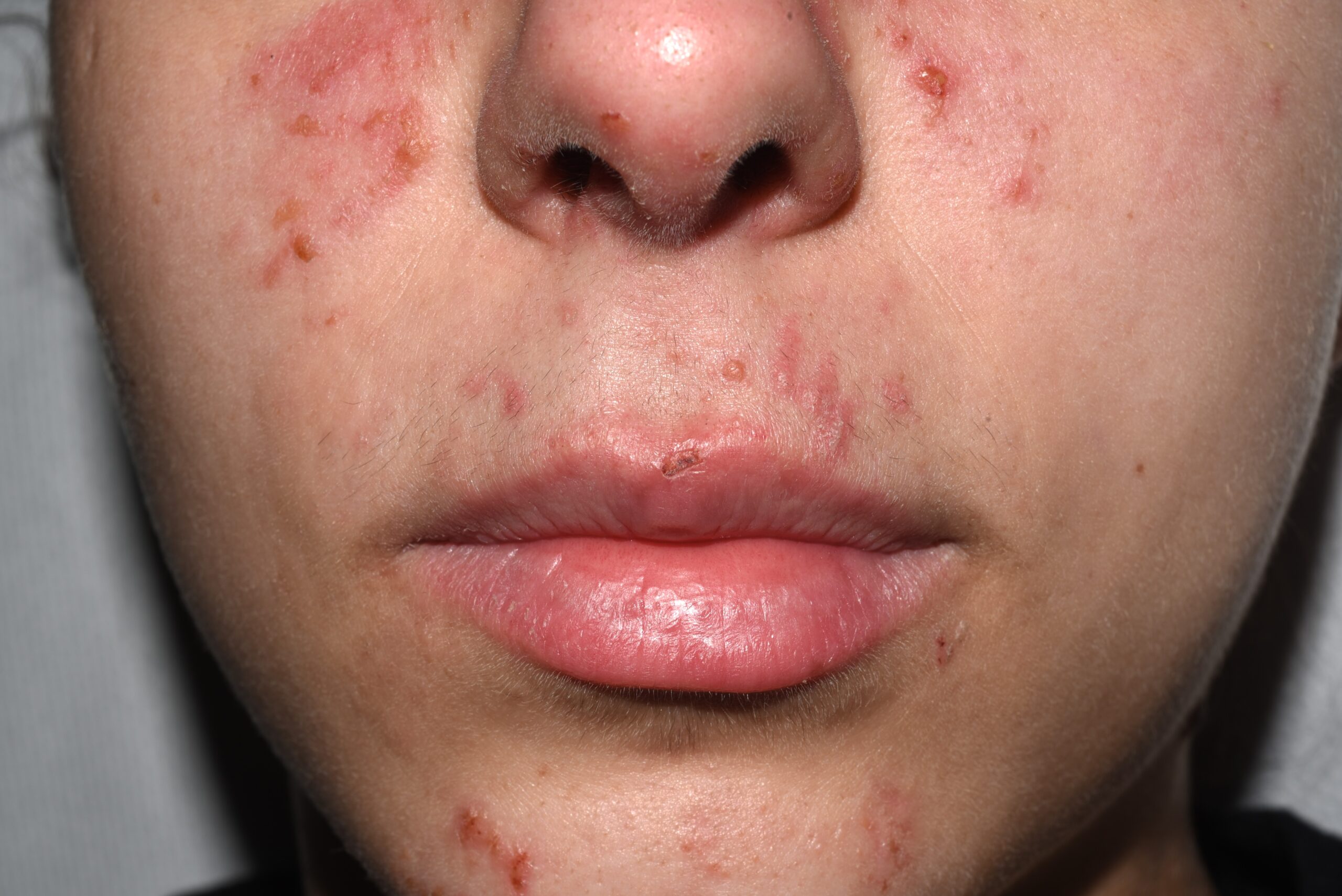
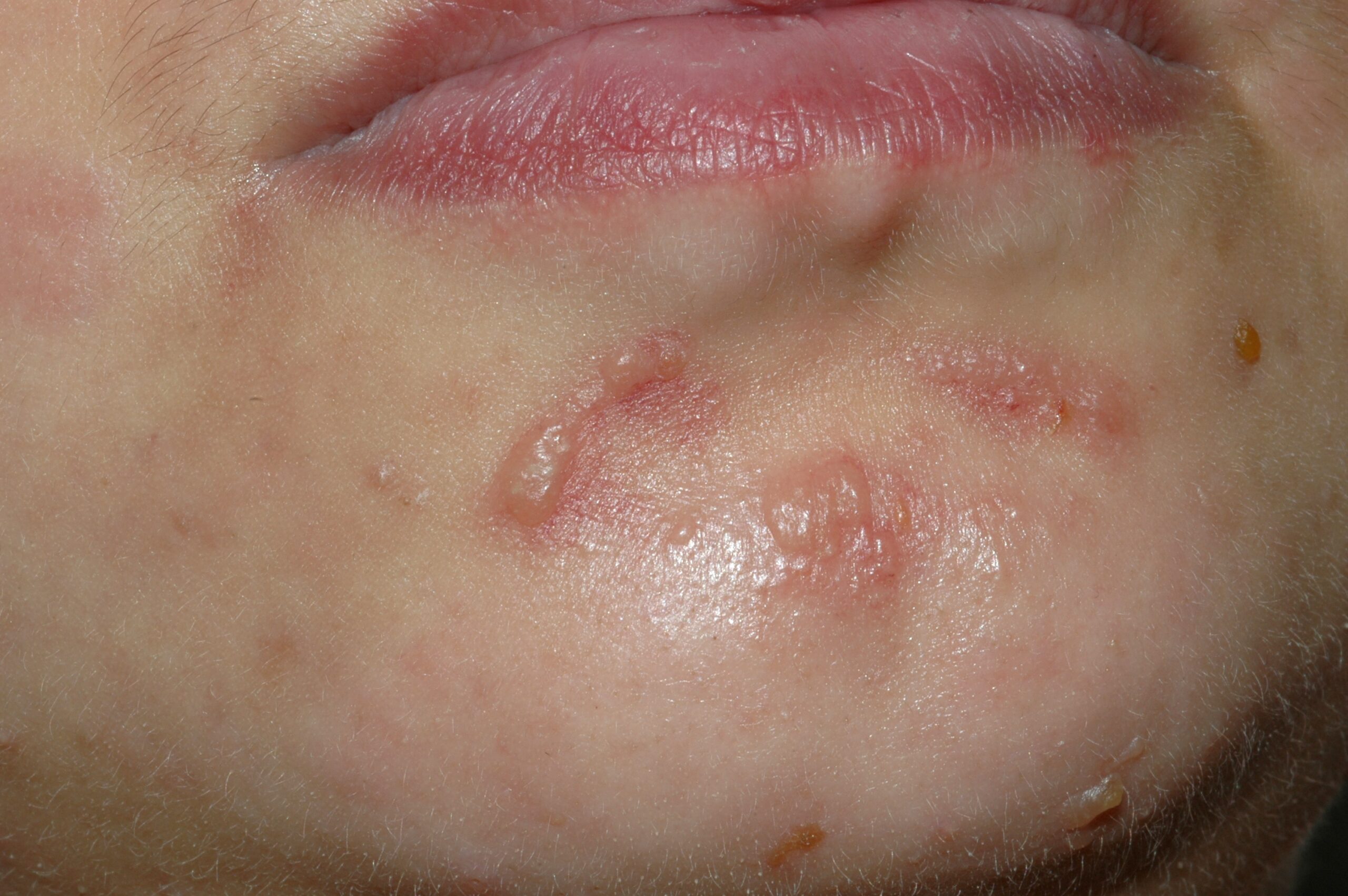
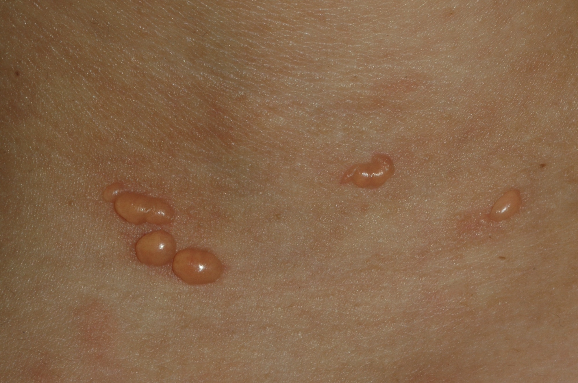
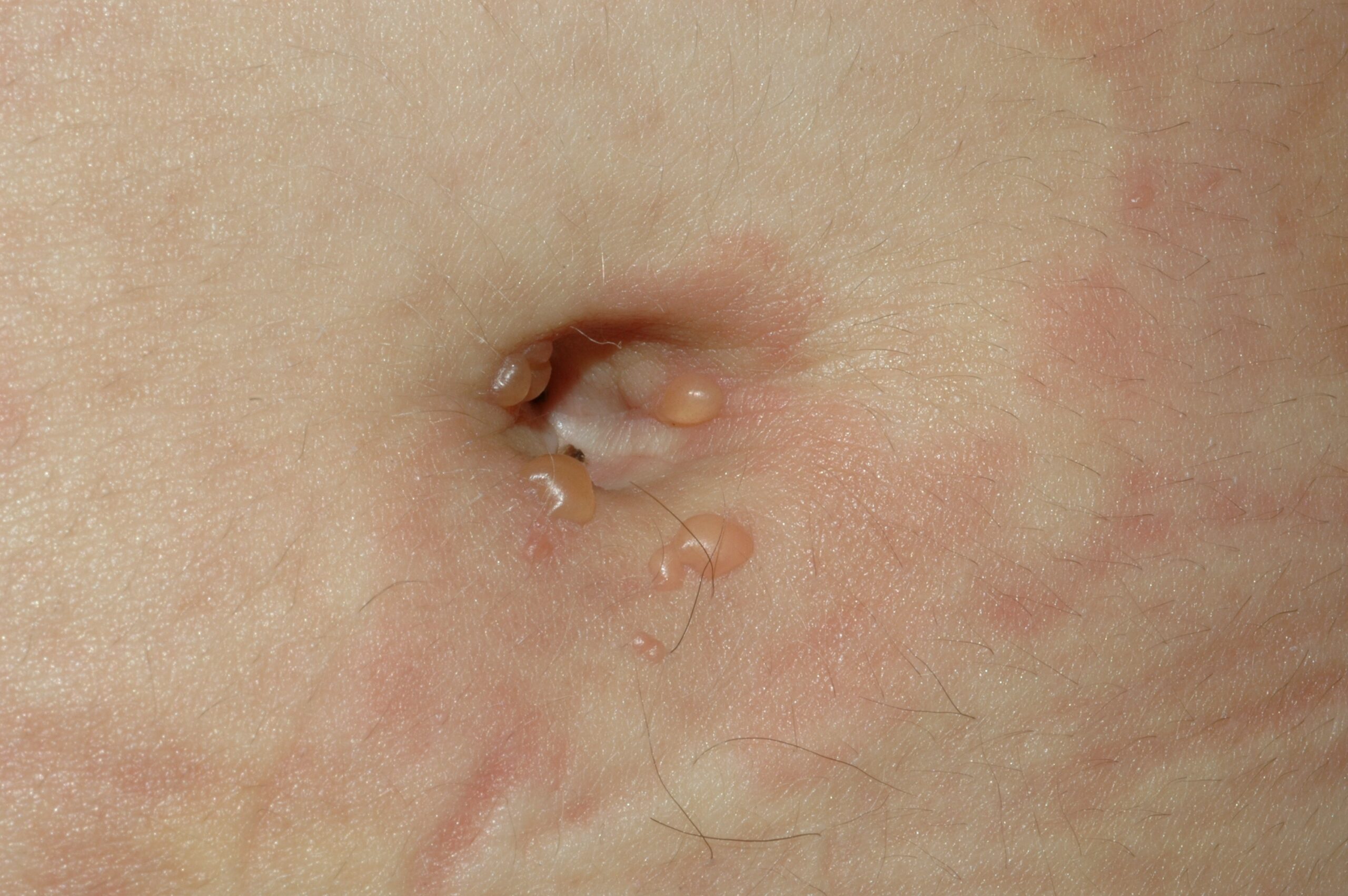
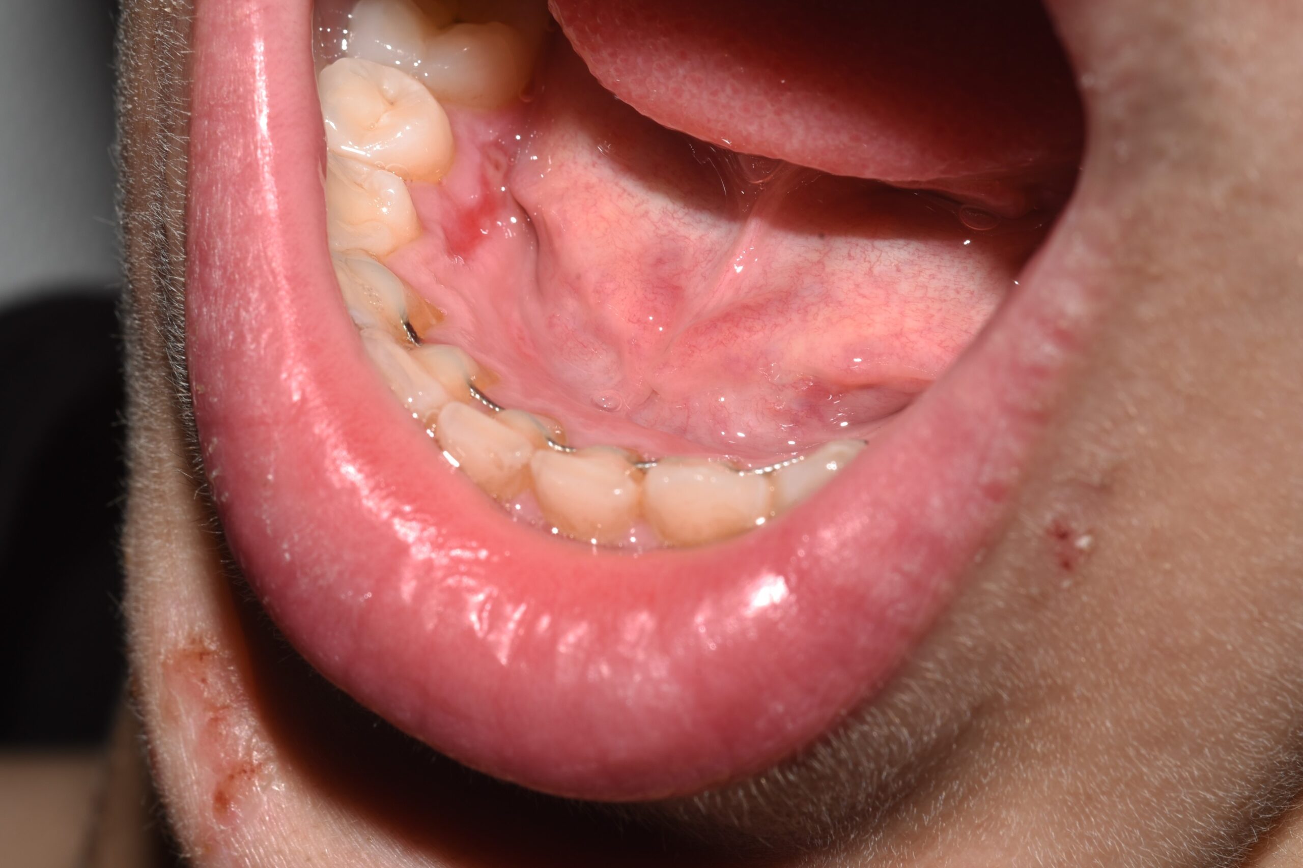
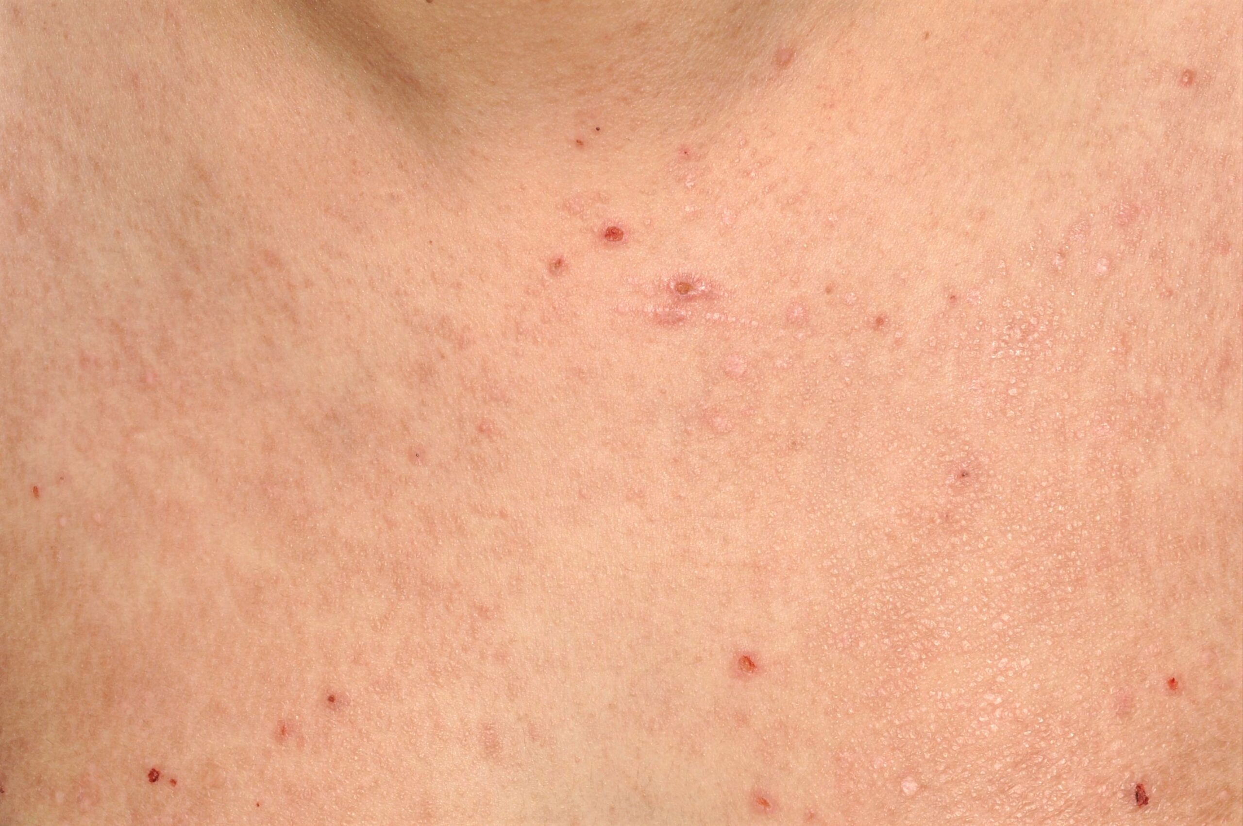
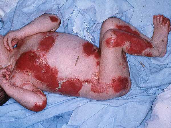
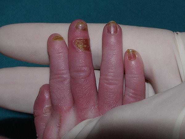
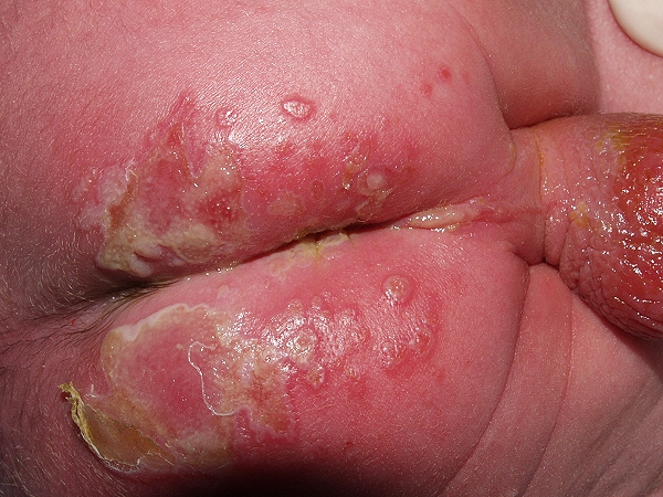
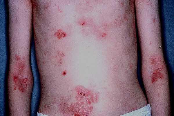
5. Bibliografía
-Larquey M, Mahé E. Érythème polymorphe postinfectieux de l’enfant. Arch Pediatr. 2016; 23:1184-1190.
-Frantz GF, McAninch SA. Mycoplasma pneumoniae–Induced Rash and Mucositis (MIRM). 2024. In: StatPearls [Internet]. Treasure Island (FL): StatPearls Publishing; 2025 Jan–.
-Heinze A, Tollefson M, Holland KE, Chiu YE. Characteristics of pediatric recurrent erythema multiforme. Pediatr Dermatol. 2018; 35:97-103.
-Saleh W, Alharbi H, Cha S. Increased prevalence of erythema multiforme in patients with COVID-19 infection or vaccination. Sci. Rep. 2024;14:2801.
-Chen N, Li M. Clinical Characteristics of 10 Children With Mycoplasma pneumoniae-Induced Rash and Mucositis. Front Pediatr. 2022;10:823376.
-Trayes KP, Love G, Studdiford JS. Erythema Multiforme: Recognition and Management. Am Fam Physician. 2019; 100:82-88.
-Tan YZ, Cakir B, Turan H. Use of valacyclovir prophylaxis in herpes virus‑associated recurrent erythema multiforme cases. Turkderm-Turk Arch Dermatol Venereol. 2022;56(2):117–119.
-Sassolas B, Haddad C, Mockenhaupt M, Dunant A, Liss Y, Bork K, Haustein UF, Vieluf D, Roujeau JC, Le Louet H. ALDEN, an algorithm for assessment of drug causality in Stevens–Johnson syndrome and toxic epidermal necrolysis: comparison with case–control analysis. Clin Pharmacol Ther. 2010;88(1):60–68.
-Hama N, Aoki S, Chen CB, Hasegawa A, Ogawa Y, Vocanson M, Asada H, Chu CY, Lan CE, Dodiuk-Gad RP, Fujiyama T, Hsieh TS, Ito K, Jerschow E, Mizukawa Y, Nakajima S, Nakamura K, Nicolas JF, Satoh TK, Shiohara T, Takahashi H, Tohyama M, Ueda T, Ura K, Watanabe H, Yamaguchi Y, Nordmann TM, Chung WH, Naisbitt D, Pincelli C, Pichler WJ, French LE, Phillips E, Abe R. Recent progress in Stevens-Johnson syndrome/toxic epidermal necrolysis: diagnostic criteria, pathogenesis and treatment. Br J Dermatol. 2024; 192(1):9-18.
-Sekula P, Dunant A, Mockenhaupt M, Naldi L, Bouwes Bavinck JN, Halevy S,
Kardaun S, Sidoroff A, Liss Y, Schumacher M, Roujeau JC; RegiSCAR study group. Comprehensive survival analysis of a cohort of patients with Stevens-Johnson syndrome and toxic epidermal necrolysis. J Invest Dermatol 2013; 133:1197.
-Bastuji-Garin S, Fouchard N, Bertocchi M, Roujeau JC, Revuz J, Wolkenstein P. SCORTEN: a severity-of-illness score for toxic epidermal necrolysis. J Invest Dermatol 2000; 115:149.
-Koh HK, Fook-Chong SMC, Lee HY. Improvement of Mortality Prognostication in Patients With Epidermal Necrolysis: The Role of Novel Inflammatory Markers and Proposed Revision of SCORTEN (Re-SCORTEN). JAMA Dermatol. 2022; 158(2):160-166.
-Nazerian A, Jafarzadeh A, Salehi S, Ghasemi M, Goodarzi A. Cyclosporin for the treatment of Stevens-Johnson syndrome (SJS) and toxic epidermal necrolysis (TEN): a systematic review. Inflammopharmacology. 2025;33(2):485-503.
-Thong BY. Drug-induced SJS/TEN: Interpreting the systematic reviews. Asia Pac Allergy. 2023; 13(2):72-76.
-Martinez Villarreal JD, Cardenas-de la Garza JA, Ionescu MA, Tatu AL, Busila C, Mokni M, Medina LMS, Jasso SMD, Poletti ED. Stevens-Johnson Syndrome and Toxic Epidermal Necrolysis: A Review of Current Management and Innovative Therapies. Int J Dermatol. 2025; 64(7):1164-1172.
-Joly P, Litrowski N. Pemphigus group (vulgaris, vegetans, foliaceus, herpetiformis, brasiliensis). Clin Dermatol. 2011; 29:432–6.
-Melchionda V, Harman KE. Pemphigus vulgaris and pemphigus foliaceus: an overview of the clinical presentation, investigations and management. Clin Exp Dermatol. 2019; 44(7):740-746.
–Popescu IA, Statescu L, Vata D, Porumb-Andrese E, Patrascu AI, Grajdeanu IA, Solovastru LG. Pemphigus vulgaris – approach and management. Exp Ther Med. 2019; 18:5056-5060.
–Harman KE, Brown D, Exton LS, Groves RW, Hampton PJ, Mohd Mustapa MF, Setterfield JF, Yesudian PD. British Association of Dermatologists’ guidelines for the management of pemphigus vulgaris 2017. Br J Dermatol. 2017;177(5):1170–1201.
-Hertl M, Jedlickova H, Karpati S, Marinovic B, Uzun S, Yayli S, Mimouni D, Borradori L, Feliciani C, Ioannides D, Joly P, Kowalewski C, Zambruno G, Zillikens D, Jonkman MF. Pemphigus. S2 guideline for diagnosis and treatment – guided by the European Dermatology Forum (EDF) in cooperation with EADV and the European Academy of Allergy and Clinical Immunology (EAACI). J Eur Acad Dermatol Venereol. 2020;34(6):1175–1193.
-Joly P, Horváth B, Patsatsi A, Uzun S, Bech R, Beissert S, Bergman R, Bernard P, Borradori L, Caproni M, Caux F, Cianchini G, Daneshpazhooh M, De D, Dmochowski M, Drenovska K, Ehrchen J, Feliciani C, Goebeler M, Groves R, Guenther C, Hofmann S, Ioannides D, Kowalewski C, Ludwig R, Lim YL, Marinovic B, Marzano AV, Mascaró JM Jr, Mimouni D, Murrell DF, Pincelli C, Squarcioni CP, Sárdy M, Setterfield J, Sprecher E, Vassileva S, Wozniak K, Yayli S, Zambruno G, Zillikens D, Hertl M, Schmidt E. Updated S2k guidelines on the management of pemphigus vulgaris and foliaceus initiated by the European Academy of Dermatology and Venereology (EADV). J Eur Acad Dermatol Venereol. 2020; 34(9):1900-1913.
-Joly P, Maho-Vaillant M, Prost-Squarcioni C, Hebert V, Houivet E, Calbo S, Caillot F Golinski ML, Labeille B, Picard-Dahan C, Paul C, Richard MA, Bouaziz JD, Duvert-Lehembre S, Bernard P, Caux F, Alexandre M, Ingen-Housz-Oro S, Vabres P, Delaporte E, Quereux G, Dupuy A, Debarbieux S, Avenel-Audran M, D’Incan M, Bedane C, Bénéton N, Jullien D, Dupin N, Misery L, Machet L, Beylot-Barry M, Dereure O, Sassolas B, Vermeulin T, Benichou J, Musette P; French study group on autoimmune bullous skin diseases. First-line rituximab combined with short-term prednisone versus prednisone alone for the treatment of pemphigus (Ritux 3): a prospective, multicentre, parallel-group, open-label randomised trial. Lancet. 2017; 389:2031-2040
-Wang HH, Liu CW, Li YC, Huang YC. Efficacy of rituximab for pemphigus: a systematic review and meta-analysis of different regimens. Acta Derm Venereol. 2015; 95:928-32.
-Mignard C, Maho-Vaillant M, Golinski ML, Balayé P, Prost-Squarcioni C, Houivet E, Calbo SB Labeille B Picard-Dahan C, Konstantinou MP, Chaby G, Richard MA, Bouaziz JD, Duvert-Lehembre S, Delaporte E, Bernard P, Caux F, Alexandre M, Ingen-Housz-Oro S, Vabres P, Quereux G, Dupuy A, Debarbieux S, Avenel-Audran M, D’Incan M, Bédane C, Bénéton N, Jullien D, Dupin N, Misery L, Machet L, Beylot-Barry M, Dereure O, Sassolas B, Benichou J, Joly P, Hébert V; French Study Group on Autoimmune Bullous Skin Diseases. Factors Associated With Short-term Relapse in Patients With Pemphigus Who Receive Rituximab as First-line Therapy: A Post Hoc Analysis of a Randomized Clinical Trial. JAMA Dermatol. 2020. doi: 10.1001/jamadermatol.2020.029.
-Bardazzi F, Loi C, Vara G, Patrizi A, Di Altobrando A. Efficacy and safety of biosimilar rituximab in the treatment of pemphigus vulgaris: a single center experience of 12 cases. J Dermatolog Treat. 2022; 33(1):580-582.
–Kimura A, Makino T, Kitayama S, Mizawa M, Ishii N, Hashimoto T, Shimizu T. Immunohistopathological analyses of a case of pemphigus vegetans with antibodies against desmoglein 1 and desmocollins 1-3. J Dermatol. 2024; 51(12):1711-1715.
–Meyer N, Misery L. Geoepidemiologic considerations of auto-immune pemphigus. Autoimmun Rev. 2010; 9:A379-82.
–Kutwin M, Kądziela M, Stein T, Kraska‑Gacka M, Woźniacka A, Żebrowska A. Senear–Usher Syndrome or Coexistence of SLE with Pemphigus Vulgaris – A Case Report with Literature Review. Journal of Clinical Medicine. 2025;14(2):409.
-Tay YE, Lee SSJ, Lim YL. Pemphigus herpetiformis with autoantibodies to desmocollins 1, 2 and 3. Clin Exp Dermatol. 2018; 43:360-62.
-Kridin K, Ahmed AR. Pemphigus herpetiformis: a systematic review of the literature. Autoimmun Rev. 2020;19(1):102427.
–Tzoumpa S, Bohelay G, Ingen-Housz-Oro S, Tancrède-Bohin E, Seta V, Joly P, Bouteiller J, Viguier MA, Berthin C, Dereure O, Lombart F, Duvert-Lehembre S, Cordel N, Mahé E, Gallais-Sérézal I, Jeudy G, Boutin D, Rousseau P, Litrowski N, Brue A, Lepelletier C, Alexandre M, Caux F, Prost C, Le Roux-Villet C. Evaluation of the efficacy and safety of dapsone alone or associated with topical corticosteroids for pemphigus foliaceus: a retrospective multicenter study. Br J Dermatol. 2025 May 22:ljaf190).
-Patel MH, Brumfiel CM, Bohrer N, Marsch A. Efficacy of rituximab in pediatric pemphigus: a literature review including pemphigus herpetiformis. JAAD Int. 2021;6:6–10.
-Tajima M, Mitsuhashi Y, Irisawa R, Amagai M, Hashimoto T, Tsuboi R. IgA pemphigus reacting exclusively to desmoglein 3. Eur J Dermatol. 2010; 20:626–9.
-Moreno AC, Santi GG, Gabbi TV, Aoki V, Hashimoto T, Maruta CW. IgA pemphigus: case series with emphasis on therapeutic response. J Am Acad of Dermatol.2014; 70:200-1.
–Svoboda SA, Huang S, Liu X, Hsu S, Motaparthi K. Paraneoplastic pemphigus: Revised diagnostic criteria based on literature analysis. J Cutan Pathol. 2021; 48(9):1133-1138.
–Zhu X, Zhang B. Paraneoplastic pemphigus. J Dermatol.2007; 34:503-11.
-Anderson HJ, Huang S, Lee JB. Paraneoplastic pemphigus/paraneoplastic autoimmune multiorgan syndrome: Part I. Clinical overview and pathophysiology. J Am Acad Dermatol. 2024; 91(1):1-10.
-Huang S, Anderson HJ, Lee JB. Paraneoplastic pemphigus/paraneoplastic autoimmune multiorgan syndrome: Part II. Diagnosis and management. J Am Acad Dermatol. 2024; 91(1):13-22.
-Barrick BJ, Lohse CM, Lehman JS. Bullous pemphigoid: Clinical features, diagnosis, and management. Mayo Clin Proc. 2021;96(9):2305–2316.
-Taghipour K, Chi C C., Bhogal B, Groves R W, Venning V, Wojnarowska F. Immunopathological characteristics of patients with bullous pemphigoid and neurological disease. J Eur Acad Dermatol Venereol. 2014; 28:569-73
-Kridin K, Bergman R. Pemphigoid and neurologic diseases: Unraveling the connection. Clin Dermatol. 2022;40(1):15–21.
-Joly P, Joly P, Roujeau JC, Benichou J, Delaporte E, D’Incan M, Dreno B, Bedane C, Sparsa A, Gorin I, Picard C, Tancrede-Bohin E, Sassolas B, Lok C, Guillaume JC, Doutre MS, Richard MA, Caux F, Prost C, Plantin P, Chosidow O, Pauwels C, Maillard H, Saiag P, Descamps V, Chevrant-Breton J, Dereure O, Hellot MF, Esteve E, Bernard P. A comparison of two regimens of topical corticosteroids in the treatment of patients with bullous pemphigoid: a multicenter randomized study. J Invest Dermatol. 2009; 129(7):1681-7.
-Sticherling M, Franke A, Aberer E, Gläser R, Hertl M, Pfeiffer C, Rzany B, Schneider S, Shimanovich I, Werfel T, Wilczek A, Zillikens D, Schmidt E. An open, multicentre, randomized clinical study in patients with bullous pemphigoid comparing methylprednisolone and azathioprine with methylprednisolone and dapsone. Br J Dermatol. 2017; 177:1299-1305).
-Lamberts A, Euverman HI, Terra JB, Jonkman MF, Horváth B. Effectiveness and Safety of Rituximab in Recalcitrant Pemphigoid Diseases. Front Immunol. 2018; 9:248.
-Polansky M, Eisenstadt R, DeGrazia T, Zhao X, Liu Y, Feldman R. Rituximab therapy in patients with bullous pemphigoid: A retrospective study of 20 patients. J Am Acad Dermatol. 2019; 81:179-186.
–Suárez Fernández R, Medina Montalvo S, Trasobares Marugán L, García Rodríguez M. Herpes gestationis tratado con ciclosporina y corticoides. Actas Dermosifiliogr. 2001; 92:302-4.
-Jenkins RE, Hern S, Black MM. Clinical features and management of 87 patients with pemphigoid gestationis. Clin Exp Dermatol. 1999; 24:255-9.
–Chan LS, Ahmed AR, Anhalt GJ, Bernauer W, Cooper KD, Elder MJ, Fine JD, Foster CS, Ghohestani R, Hashimoto T, Hoang-Xuan T, Kirtschig G, Korman NJ, Lightman S, Lozada-Nur F, Marinkovich MP, Mondino BJ, Prost-Squarcioni C, Rogers RS 3rd, Setterfield JF, West DP, Wojnarowska F, Woodley DT, Yancey KB, Zillikens D, Zone JJ. The first international consensus on mucous membrane pemphigoid: definition, diagnostic criteria, pathogenic factors, medical treatment, and prognostic indicators. Arch Dermatol. 2002; 138(3):370-379.
-Kridin K. Mucous membrane pemphigoid: a systematic review of epidemiology, clinical characteristics, and management. J Am Acad Dermatol. 2018;78(3):563–571.
–Bernard P, Antonicelli F. Mucous membrane pemphigoid: treatment update and future directions. Front Med (Lausanne). 2021; 8:730651.
-Tan WH, Sim J, Sultana R, Lee HY. Rituximab in Subepidermal Blistering Diseases. Dermatology. 2023; 239(1):5-11.
-Kridin K. Subepidermal autoimmune bullous diseases: overview, epidemiology, and associations. Immunol Res. 2018; 661:6–17.
-Sankari H, Hietikko M, Kurppa K, Kaukinen K, Mansikka E, Huhtala H, Laurila K, Reunala T, Hervonen K, Salmi T, Lindfors K. Intestinal TG3- and TG2-Specific Plasma Cell Responses in Dermatitis Herpetiformis Patients Undergoing a Gluten Challenge. Nutrients. 2020; doi: 10.3390/nu12020467.
–Lebwohl B, Sanders DS, Green PHR. Coeliac disease. Lancet. 2018; 391:70-81.
-King JA, Jeong J, Underwood FE, Quan J, Panaccione N, Windsor JW, Coward S, deBruyn J, Ronksley PE, Shaheen AA, Quan H, Godley J, Veldhuyzen van Zanten S, Lebwohl B, Ng SC, Ludvigsson JF, Kaplan GG. Incidence of Celiac Disease Is Increasing Over Time: A Systematic Review and Meta-analysis. Am J Gastroenterol. 2020; 115:507-525.
-Salmi TT. Dermatitis herpetiformis. Clin Exp Dermatol. 2019; 44:728-731.
–Reunala T, Salmi TT, Hervonen K, Kaukinen K, Collin P. Dermatitis Herpetiformis: A Common Extraintestinal Manifestation of Coeliac Disease. Nutrients. 2018; doi: 10.3390/nu10050602.
-Salmi TT, Hervonen K, Kurppa K, Collin P, Kaukinen K, Reunala T. Celiac disease evolving into dermatitis herpetiformis in patients adhering to normal or gluten-free diet. Scand J Gastroenterol. 2015; 50:387-92.
-Salmi TT, Hervonen K, Kautiainen H, Collin P, Reunala T. Prevalence and incidence of dermatitis herpetiformis: a 40-year prospective study from Finland. Br J Dermatol. 2011; 165(2):354-9.
-West J, Fleming KM, Taj L, Tata LJ, Crooks CJ, Card TR. Incidence and prevalence of coeliac disease and dermatitis herpetiformis in the UK over two decades: population‑based study. Am J Gastroenterol. 2014; 109(5):757–768.
–Cardones AR, Hall RP 3rd. Management of dermatitis herpetiformis. Immunol Allergy Clin North Am. 2012; 32:275-81.
–Al-Toma A, Volta U, Auricchio R, Castillejo G, Sanders DS, Cellier C, Mulder CJ, Lundin KEA. European Society for the Study of Coeliac Disease (ESsCD) guideline for coeliac disease and other gluten-related disorders. United European Gastroenterol J. 2019; 7:583-613.
–Mansikka E, Hervonen K, Kaukinen K, Ilus T, Oksanen P, Lindfors K, Laurila K, Hietikko M, Taavela J, Jernman J, Saavalainen P, Reunala T, Salmi T. Gluten Challenge Induces Skin and Small Bowel Relapse in Long-Term Gluten-Free Diet-Treated Dermatitis Herpetiformis. J Invest Dermatol. 2019; 139:2108-2114.
-Ghaoui N, Hanna E, Abbas O, Kibbi AG, Kurban M. Update on the use of dapsone in dermatology. Int J Dermatol. 2020. doi: 10.1111/ijd.14761.
-Casanova JM, Cortada JL, Puig T, Rubio M, Ramos J. Lupus eritematoso cutáneo subagudo. Tratamiento con sulfona. Med Cutan Ibero Lat Am. 1987; 15:259-263.
–Estève C, Samson M, Guilhem A, Nicolas B, Leguy-Seguin V, Berthier S, Bonnotte B, Audia S. Eficacy and safety of dapsone as second line therapy for adult immune thrombocytopenia: A retrospective study of 42 patients. PLoS One. 2017; 12:e0187296. doi: 10.1371/journal.pone.0187296.
-Lammer J, Hein R, Roenneberg S, Biedermann T, Volz T. Drug-induced Linear IgA Bullous Dermatosis: A Case Report and Review of the Literature. Acta Derm Venereol. 2019; 99:508-515.
-Saleem M, Iftikhar H. Linear IgA Disease: A Rare Complication of Vancomycin. Cureus. 2019; 11:e4848. doi: 10.7759/cureus.4848.
-Saleem M, Arif T. Drug-induced linear IgA bullous dermatosis. J Pharmacol Pharmacother. 2019; 10(2):62–66.
-Vives P, Casanova JM, Hernanz JM, Lecona M, Jaqueti G. Dermatosis ampollosa crónica benigna infantil. Actas Dermosifiliogr. 1984; 75:55-61.
-Sanmartín V, Baradad M, Egido R, Casanova JM. Dermatosis ampollosa crónica benigna infantil (dermatosis IgA lineal de la infancia) Piel. 2009; 24(8):457-61.
-Juratli HA, Sárdy M. Lineare IgA-Dermatose. Hautarzt. 2019; 70:254-259.
-Zone JJ, Taylor TB, Kadunce DP, Chorzelski TP, Schachner LA, Huff JC, Meyer LJ, Petersen MJ. IgA antibodies in chronic bullous disease of childhood react with 97 kDa basement membrane zone protein. J Invest Dermatol. 1996; 106:1277-80
-Fortuna G, Marinkovich MP. Linear immunoglobulin A bullous dermatosis. Clin Dermatol. 2012; 30:38-50.
-Koga H, Teye K, Yamashita K, Ishii N, Tsuruta D, Nakama T. Detection of anti-type VII collagen IgE antibodies in epidermolysis bullosa acquisita. Br J Dermatol. 2019; 180(5):1107-1113.
–Iranzo P, Herrero-González JE, Mascaró-Galy JM, Suárez-Fernández R, España A. Epidermolysis bullosa acquisita: a retrospective analysis of 12 patients evaluated in four tertiary hospitals in Spain. Br J Dermatol. 2014; 171(5):1022-30).
–Mehren CR, Gniadecki R. Epidermolysis bullosa acquisita: current diagnosis and therapy. Dermatol Reports. 2011; 3(3):e38.
–Vorobyev A, Ludwig RJ, Schmidt E. Epidermolysis bullosa acquisita: current and emerging treatment options. Am J Clin Dermatol. 2021;22(3):371–390.
–de Risi-Pugliese T, Cohen Aubart F, Haroche J, Moguelet P, Grootenboer-Mignot S, Mathian A, Ingen-Housz-Oro S, Hie M, Wendremaire N, Aucouturier F, Lepelletier F, Miyara M, Bader-Meunier B, Rémy P, Fabien N, Francès C, Barete S, Amoura Z. Clinical, histological, immunological presentations and outcomes of bullous systemic lupus erythematosus: 10 New cases and a literature review of 118 cases. Semin Arthritis Rheum. 2018; 48:83-89.
-Chanprapaph K, Sawatwarakul S, Vachiramon V. A 12-year retrospective review of bullous systemic lupus erythematosus in cutaneous and systemic lupus erythematosus patients. Lupus. 2017; 26:1278-1284.
–Sprow G, Afarideh M, Dan J, Hedberg ML, Werth VP. Bullous systemic lupus erythematosus in females. Int J Womens Dermatol. 2022; 8(3):e034.
–Duan L, Chen L, Zhong S, Wang Y, Huang Y, He Y, Chen J, Shi G. Treatment of Bullous Systemic Lupus Erythematosus. J Immunol Res. 2015; 2015:167064.
–Lowe CD, Brahe CA, Green B, Lam TK, Meyerle JH. Bullous systemic lupus erythematosus successfully treated with rituximab. Cutis. 2019; 103(6):E5-E7.
–Hübner F, Recke A, Zillikens D, Linder R, Schmidt E. Prevalence and age distribution of subepidermal autoimmune bullous disorders in Germany. J Invest Dermatol. 2019;139(2):293–298.
-Hübner F, Langan EA, Recke A. Lichen Planus Pemphigoides: From Lichenoid Inflammation to Autoantibody-Mediated Blistering. Front Immunol. 2019; 10:1389.
-Zaraa I, Mahfoudh A, Sellami MK, Chelly I, El Euch D, Zitouna M, Mokni M, Makni S, Ben Osman A. Lichen planus pemphigoides: four new cases and a review of the literature. Int. J. Dermatol. 2013; 52:406-12.
–Mithra S, Parimalam K, Sowmiya R. Comparative study of the efficacy of azathioprine, dapsone, and NB‑UVB phototherapy as steroid‑sparing modalities in generalized lichen planus. Indian J Dermatol Venereol Leprol. 2025; 91:59-64.
-Laimer M, Prodinger C, Bauer JW. Hereditary epidermolysis bullosa. J Dtsch Dermatol Ges. 2015; 13:1125–1133.
–Watkins J. Diagnosis, treatment and management of epidermolysis bullosa. Br J Nurs. 2016; 25:428-431.
–Khani P, Ghazi F, Zekri A, Nasri F, Behrangi E, Aghdam AM, Mirzaei H. Keratins and epidermolysis bullosa simplex. J Cell Physiol. 2018; 234:289-297.
-Casanova JM, Martinez P, Vega M, Gomá AR, Moreno A. Epidermolisis ampollosa herpetiforme (Dowling-Meara). Actas Dermo-Sif 1987; 78:117-120.
-Lincoln V, Pillay K, Wessels Q. Herlitz junctional epidermolysis bullosa: a clinical review. Curationis. 2018;41(1):a1849.
-Casanova JM, González-Herrada CM, Frías J, Lázaro P, Lecona M, Cervera A. Epidermolisis ampollosa juntural tipo Herlitz. Actas Dermo-Sif 1984; 75:332-336.
-Yang CS, Kroshinksy D, Cummings BM. Neonatal junctional epidermolysis bullosa: treatment conundrums and ethical decision making. Am J Clin Dermatol. 2014; 15:445-50.
–Cianfarani F, Zambruno G, Castiglia D, Odorisio T. Pathomechanisms of Altered Wound Healing in Recessive Dystrophic Epidermolysis Bullosa. Am J Pathol. 2017; 187:1445-1453).
–Li AW, Prindaville B, Bateman ST, Gibson TE, Wiss K. Inpatient management of children with recessive dystrophic epidermolysis bullosa: A review. Pediatr Dermatol. 2017; 34:647-655).
-Micaroni M, Giacchetti G, Plebani R, Xiao GG, Federici L. ATP2C1 gene mutations in Hailey-Hailey disease and possible roles of SPCA1 isoforms in membrane trafficking. Cell Death Dis. 2016; 7:e2259.
-Engin B, Kutlubay Z, Çelik U, Serdaroğlu S, Tüzün Y. Hailey-Hailey disease: A fold (intertriginous) dermatosis. Clin Dermatol. 2015; 33:452-5.
-Patel VM, Rubins S, Schwartz RA, Septe M, Rubins A. Hailey-Hailey disease: a diagnostic challenge. Cutis. 2019; 103:157-159.
-Oliveira A, Arzberger E, Pimentel B, de Sousa VC, Leal-Filipe P. Dermoscopic and reflectance confocal microscopic presentation of Hailey-Hailey disease: A case series. Skin Res Technol. 2018; 24:85-92.
-Lapa T, Breslavets M. Treatment of Hailey-Hailey disease with narrowband phototherapy and acitretin: A case report. SAGE Open Med Case Rep. 2019; 7:2050313X19845221.
-Albers LN, Arbiser JL, Feldman RJ. Treatment of Hailey-Hailey Disease With Low-Dose Naltrexone. JAMA Dermatol. 2017; 153:1018-1020.